Download
1 / 49
660 likes | 1.16k Vues
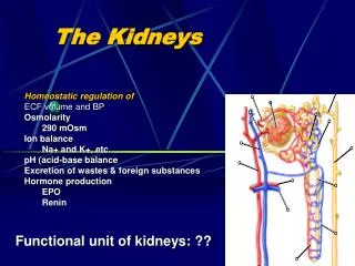
Glomerular filtration, renal blood flow and their control. Functions of the Kidneys. T he K idneys. Perfect examples of homeostatic organs Every day they filter gallons of fluid from the blood stream They then process the filtrate allowing wastes and excess ions to leave the body

E N D
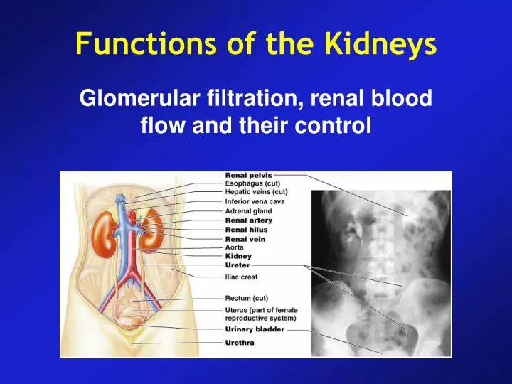
Glomerular filtration, renal blood flow and their control Functions of the Kidneys
The Kidneys • Perfect examples of homeostatic organs • Every day they filter gallons of fluid from the blood stream • They then process the filtrate allowing wastes and excess ions to leave the body • Role of the lungs and skin in excretion...
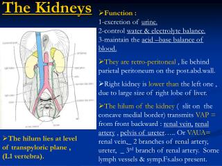
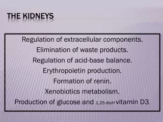
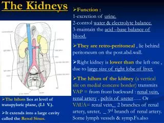
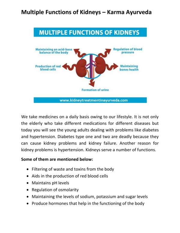
Functions of the Kidneys • Excretion of metabolic waste products, foreign chemicals, drugs and hormone metabolites • Regulation of water and electrolyte balances • Regulation of arterial pressure • Regulation of acid-base balance • Secretion, metabolism and excretion of hormones • Gluconeogenesis
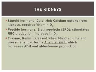
Functions of the Kidneys • Regulatory functions • Renin in regulation of blood pressure • Erythropoietin in stimulation of red blood cell production • Conversion of Vit D to its active form “1,25-dihydroxyvitamin D”
Renal Failure and Hemodialysis • Renal failure (infections, crash, toxicants, chronic hypertension or glomerulonephritis) • Symptoms: uremia, diarrhea, vomiting, labored breathing, convulsions, coma ... • Signs become obvious only after 75% of the renal function has been lost • Uremia occurs after 90% of the nephrons have been damaged • Hemodialysis (3 times/week) • In case of non-reversible damage; kidney transplant
Kidneys Urinary Tract • Organs that excrete urine • Organs that eliminate urine: • ureters (paired tubes) • urinary bladder (muscular sac) • urethra (exit tube) Urination or Micturition • Process of eliminating urine • Contraction of muscular urinary bladder forces urine through urethra, and out of body
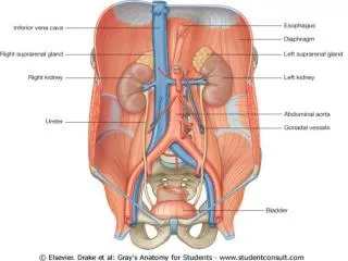
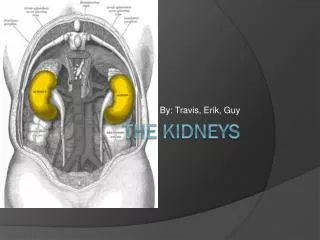
Location and Structure • Adrenal glands as separate organs
Hilus • Entry for renal artery and renal nerves • Exit for renal veins and ureter
Typical Adult Kidney • Is about 12 cm long, 6 cm wide, and 3 cm thick • Weighs about 150 g • Is protected and stabilized by 3 concentric layers of connective tissue: • Renal capsule • Adipose capsule • 3. Renal fascia
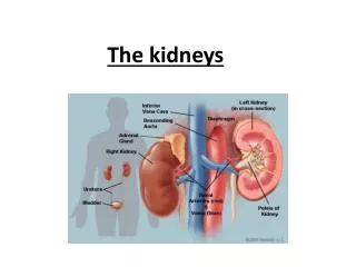
Superficial outer cortex and inner medulla • The medulla consists of 6-18 renal pyramids; tip of each pyramid is known as the renal papilla which projects into the renal sinus • Adjacent renal pyramids are separated by renal columns, which extend into the medulla • The cortex of each renal lobe (which has a renal pyramid, overlying renal cortex, and adjacent tissues of the renal columns) is composed of roughly 1.25 million nephrons where urine production begins • Ducts within each renal papilla discharge urine into a cup-shaped drain called a minor calyx • Four or five minor calyces merge to form a major calyx • Two or three major calyces combine to form the renal pelvis, a large funnel shaped chamber • Renal pelvis, which fills most of the renal sinus, is connected to the ureter, which drains the kidney
Blood supply to the kidneys • Each kidney receives blood from a renal artery • A Renal artery originates along lateral surface of abdominal aorta near level of the superior mesenteric artery • Renal artery supplies blood to the segmental arteries • Segmental arteries further divide into a series of interlobar arteries • Interlobar arteries radiate outward through renal columns between renal pyramids • Interlobar arteries supply blood to the arcuate arteries • Arcuate arteries arch along boundary between cortex and medulla if kidney • Arcuate arteries give rise to a number of interlobular arteries, which supply cortical portions of adjacent renal lobes • Branching from each interlobular artery are numerous afferent arterioles • Afferent arterioles deliver blood to the capillaries supplying individual nephrons • Renal venules follow similar opposing pattern ending with renal veins
The Blood Supply to the Kidneys (C). A flowchart of renal circulation. (D). Blood flow to the nephron
Glomerular and Peritubular Capillary Beds
Functional unit of the kidney: the nephron • Total of about 2.5 million in the 2 kidneys. • Each nephron consists of 2 functional components: • The tubular component (contains what will eventually become urine) • The vascular component (blood supply) • The mechanisms by which kidneys perform their functions depends upon the relationship between these two components.
Juxtamedullary Nephrons • 15% of nephrons • Have long loops of Henle that extend deep into medulla
Micturition • Micturition is the process by which the urinary bladder empties when it becomes filled • Bladder fills progressively until the tension in its walls rises above a threshold • This elicits the second step, a nervous reflex called micturition reflex that empties the bladder • Although it is an autonomic spinal cord reflex, it can also be inhibited or facilitated by centers in the cerebral cortex or brain stem
Anatomy and Nervous Connections of the Bladder • The body and the neck • Detrusor muscle • Trigone • Internal and external sphincters
Ureters and Transport of Urine • Renal calyces, ureters and urinary bladder • Entrance of ureters into the bladder at the trigone region • Vesicoureteral reflux • Pain sensation in the ureters • Ureters are well supplied with pain nerve fibers • When ureters become blocked, intense reflex constriction occurs with severe pain • Uretero-renal reflex & prevention of excessive fluid flow
Micturition reflex and urination • Urination coordinated by micturition reflex • Micturition = urination • Ordinarily, it continues to collect urine until about 200 ml accumulated • Initiated by stretch receptors in wall of bladder • Impulses transmitted to sacral region of the spinal cord and then back to the bladder via the pelvic splanchinic nerves • Urination requires coupling micturition reflex with relaxation of external urethral sphincter • When one chooses not to void, the reflex contractions of the bladder will stop within a minute or so and urine will continue to accumulate in the bladder.
The Micturition Reflex Components of the reflex arc that stimulates smooth muscle contractions in the urinary bladder. Micturition occurs after voluntary relaxation of the external urethral sphincter.
Incontinence and Urinary retention • Incontinence: being unable to voluntarily control the external sphincter • Normal in children under 2 years of age • May happen in older children • Emotional problems, pressure, or nervous system problems (spinal cord injury or stroke) • Urinary retention: A condition that the bladder is unable to expel its contained urine. • General anesthesia • In elderly men, hypertrophy of the prostate gland. Its enlargement narrows the urethra and make it difficult to void
Urine Formation a) Glomerular Filtration b) Reabsorption c) Secretion Urinary excretion rate = Filtration – Reabsorption + Secretion
The Renal Corpuscle • Each renal corpuscle: • is 150–250 µm in diameter • includes Glomerulus andBowman’s capsule
Glomerular Filtration • Composition of glomerular filtrate • Similarity to plasma • No proteins • Exceptions: calcium and fatty acids • They are partially bound to plasma proteins • GFR is about 20% of the renal plasma flow • GFR is determined by the balance between hydrostatic and oncotic forces acting across the capillary membrane • Normally GFR is about 125 ml/min or 180 L/day
Glomerular Capillary Membrane • It has 3 (instead of 2 the usual 2) major layers: • Endothelium of the capillary • A basement membrane • A layer of epithelial cells (podocytes) • The capillary endothelium is perforated by thousands of small holes (fenestrae)
Glomerular Filtration • Filterebility of solutes is inverselyrelatedtotheir size Negatively charged molecules (such as plasma proteins) are less filtered
Determinants of GFR • Forces favoring filtration • Glomerular hydrostatic pressure : 60 mmHg • Bowman’s capsule oncotic pressure : 0 mmHg • Forces opposing filtration • Bowman’s capsule hydrostatic pressure : 18 mmHg • Glomerular capillary oncotic pressure: 32 mmHg
Glomerular Hydrostatic Pressure • It is determined by 3 variables 1) Arterial pressure 2) Afferent arteriolar resistance 3) Efferent arteriolar resistance • Constriction of afferent arterioles reduces GFR • Effect of efferent arteriolar constriction depends on the severity of constriction • Modest efferent constriction increases GFR • But severe constriction tends to reduce GFR
Blood Supply to the Kidney • Kidneys receive 20–25% of total cardiac output • 1200 ml of blood flows through kidneys each minute
Determinants of Renal Blood Flow • Oxygen supply & Metabolic rate of kidneys • Oxygen delivered to the kidneys far exceeds their metabolic needs • Renal vascular resistance: • Interlobular arteries • Afferent arterioles • Efferent arterioles • GFR is relatively constant over an arterial pressure of 80 to 170 mmHg (autoregulation) • Blood flow in the vasa recta Renal Blood Flow = Renal artery pressure – renal vein pressure / Total renal vascular resistance
Physiologic Control of Glomerular Filtration and Renal Blood Flow • Sympathetic activation can constrict renal arterioles and reduces blood flow and thus decreases GFR • Hormonal and Autocoid Control of Renal Circulation • Noradrenaline, Adrenaline and Endothelin constrict renal vessels and decrease GFR • Angiotensin II constricts efferent arterioles • EDRF or Nitric Oxide decreases renal vascular resistance and increases GFR • Prostaglandins and bradykinin tend to increase GFR
Autoregulation of GFR and Renal Blood Flow • The relative constancy of GFR and renal blood flow is referred to as “autoregulation” • Importance of autoregulation in preventing extreme changes in renal excretion • Normal GFR : 180 L/day • Tubular reabsorption : 178.5 L/day • “Glomerulotubular balance” • Even with these special control mechanisms, changes in arterial pressure still have significant effects on water and solute excretion: • Pressure diuresis and natriuresis
Tubuloglomerular Feedback Mechanism and Autoregulation of GFR • The kidneys have a feedback mechanism that links changes in NaCl concentration at the macula densa with the control of renal arteriolar resistance • The juxtaglomerular complex: • Macula densa and • Juxtaglomerular cells
Tubuloglomerular Feedback Mechanism and Autoregulation of GFR