Download
1 / 54
1.26k likes | 4k Vues
Inborn Errors of Metabolism. Dr B.Vahabi. Lecture outcomes. Understand the general pathophysiology underlying the inborn errors of metabolism (IEMs) Review some important IEMs Understand the genetic inheritance of IEMs Review the general diagnostic methods used for detection of IEMs

E N D
Inborn Errors of Metabolism Dr B.Vahabi
Lecture outcomes • Understand the general pathophysiology underlying the inborn errors of metabolism (IEMs) • Review some important IEMs • Understand the genetic inheritance of IEMs • Review the general diagnostic methods used for detection of IEMs • Discuss the current treatment options for people suffering from IEMs
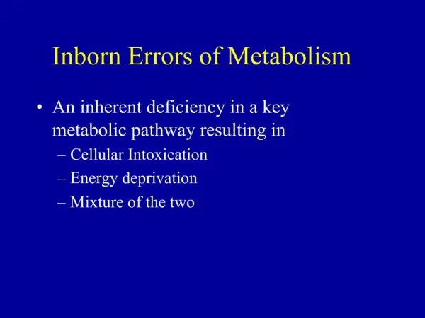
Metabolism • Metabolism is the sum of all the chemical reactions in the body • Some chemical reactions are involved in breaking down molecules, others are involved in building up (synthesis) • A metabolic pathway consists of SEVERAL STAGES involved in the conversion of one metabolite to another.
Metabolism Food Enzyme A Amino acids Carbohydrates Lipids Nucleic acids Enzyme B Protein Carriers Energy Biomolecules
Errors in Metabolism • If an error occurs in the gene that codes for the enzyme a FAULT occurs. • This fault is caused by a mutation in the genetic code. • Subsequently the enzyme is not produced and the pathway breaks down. • These are called INBORN ERRORS OFMETABOLISM.
Metabolite D Enzyme 1 Enzyme 2 Metabolite A Metabolite B Metabolite C Accumulation of substances present in small amount Deficiency of specific final products Deficiency of critical intermediary products
The concept of inborn errors of metabolism (IEM) was first introduced by Archibald Garrod in 1908.
Incidence • More than 300 human diseases are known today that are caused by IEM • Overall prevalence of 1 in 5000 • However the prevalence of each disease has many variables • Certain IEMs have a race related prevalence • e.g Tay-Sachs in Ashkenazi Jews
Inheritance • Majority IEMs are autosomal recessive • Some IEMs are X-linked (Mothers are carriers) • Mitochondrial diseases have also been detected
Usual clinical presentation of IEMs • Young Children • Recurring vomiting • Dysmorphic features (characteristic facial expression, slant of eyes) • Developmental delay (milestones) • Seizures • Mental retardation • Neonates • Poor feeding • Vomiting • Apnoea (breathing disorder) • Irritability • Abnormal tone • Seizures
Categories of IEMs • Amino acid metabolism disorders • Carbohydrate metabolism disorders • Lysosomal storage disorders • Fatty acid oxidation disorders • Urea cycle defects • Peroxisomal disorders • Mitochondrial disorders
Amino acid metabolism disorders • A heterogeneous group of disorders • Block at early step of metabolic pathway resulting in accumulation of amino acids • Block at later stages of metabolic pathway resulting in accumulation of metabolites • Defect in transport mechanism of amino acids resulting in decreased intestinal transport and increased urinary excretion
Amino acid metabolism disorders Examples: • Phenylketonuria- phenylalanine • Homocysteinuria- methionine • Maple syrup urine disease- Leucine, isoleuscine and valine • Tyrosinaemia- Tyrosine
Phenyketonuria (PKU) • Most prevalent disorder caused by inborn errors of amino acid metabolism • Caused by mutations in phenyalanine hydroxylase (PAH) gene • PAH converts phenyalanine into tyrosine and requires the cofactor tetrahydrobiopterin (BH4), molecular oxygen and iron • Loss of PAH activity increased concentrations of phenyalanine in blood an d toxic concentrations in the brain
Molecular genetics and classification • The PAH gene consists of 13 exons • PKU arises when both alleles are mutated (548 separate mutations) • Some mutations only partly inhibit the enzyme activity mild PKU • About 1-2% of cases of PKU are due to mutations in genes coding for enzymes involved in BH4 biosynthesis
Pathophysiology of PKU • Phenylalanine’s entry into the brain is mediated by the large neutral aminoacid carrier L-aminoacid transporter (LAT1) • Raised phenylalanine concentration can induce damage in the brain by: • Reducing formation of myeline in brain white matter • Inhibition of LAT1 carriers and neutral amino acids from entering the brain • Reduced activity of pyruvate kinase • Disturbed glutamatergic neurotransmission • Reduced activity of the enzyme 3-hydroxy-3-methylglutaryl coenzyme reductase.
Presentation of PKU • Developmental Delay • Musty odour • Mental Retardation (decreased Myelin formation) • Epilepsy • Autism • Hypopigmentation (decreased melanin) • Blood phenylalanine concentrations >1200µMol/Lit • Detected by newborn screening (heel prick test) • Can be dietary controlled.
Carbohydrate metabolism disorders • A heterogeneous group of disorders • Caused by inability to metabolize specific sugars, aberrant glycogen synthesis or disorders of gluconeogenesis • Manifest with hypoglycemia, hepatosplenomegaly, lactic acidosis or ketosis
Carbohydrate metabolism disorders Examples: • Glycogen storage diseases • Galactosemia • Fructose intolerance • Fructose 1,6-diphosphate deficiency
Glycogen storage diseases (GSDs) • Characterized by abnormal inherited glycogen metabolism in the liver, muscle and brain. • Lead to build up of glycogen in tissues • Categorised numerically (0-X) (e.g. Type II, Type III etc.)
Von Gierke disease (GSD type I) • Caused by defective liver glucose 6-phosphatase activity • Mutations can either be in: • Gene coding for the liver glucose-6-phosphatase • Gene coding for endoplasmic reticulum substrate • Product transport proteins of the glucose-6-phosphatase system
Presentation of Von Gierke 1a disease • Initial symptoms are due to hypoglycaemia and include: • Tremor, irritability, hyperventilation, apnea, convulsions, paleness, sweating, cerebral edema, coma and death • Older infants may present with: • Doll-like facial appearance, frequent lethargy, difficult arousal from sleep, overwhelming hunger, protuberant abdomen, relatively thin extremities. • With ageing the patient presents: • Poor growth, short stature, and rachitic changes • Most striking laboratory findings: • Hypoglycaemia, lactic acidosis, hyperlipidemia, hyperuricaemia, mosaic pattern of the liver, pale staining of the tissue and swollen hepatocytes
Presentation of Von Gierke 1b disease In addition to clinical symptoms seen in GSD-1a: Recurrent infections Neutropenia Neutrophil dysfunction Inflammatory bowl disease Fever Diarrhea Perioral and anal ulcers
Lysosomal storage disorders • Are caused by accumulation of glycoproteins, glycolipids or glycosaminoglycans within lysosomes in various tissues • Usually present later in infancy with organomegaly, facial coarseness and neurodegeneration • Show progressively degenerative course
Lysosomal storage disorders Examples • Tay-Sachs • Niemann-Pick disease • Gaucher’s disease
Tay-Sachs disease • Lack of lysosomal β-hexosaminidase A (Hex-A) enzyme activity • Hex-A is responsible for production of hexosaminidase A • Hexosamindase A breaks down a fatty acid substance called GM2 ganglioside in nerve cells • Accumulation of GM2-ganglioside has a toxic effect on cells neuronal deterioration mental and motor retardation
Presentation of Tay-Sachs disease • Infant appears normal at birth but within few weeks may become less visually attentive, hypotonic and easily startled by sound, light or touch • By 6-8 months developmental delay becomes obvious • Fundiscopic examination of retina reveals a whitish surrounding lipid deposition • By 1 year marked reduction in purposeful movement, child becomes spastic and lethargic • Vision deteriorates • Frequent seizures • By age 2 years the child is in a vegetative state and requires constant care • Feeding difficulty • A light cherry red spot in the middle of the eye • The brain increases in weight and size but shows generalized atrophy and reduction in nerves and white matter • Deafness • Usually death before age of 5.
Diagnosis and Management There are 3 important steps in the diagnosis and management of IEM: • Suspicion • Evaluation • Treatment
Suspicion • An important key to diagnosing IEM is thinking about the possibility in the first place • The symptoms are very common and non-specific • Screening allows for the differential diagnosis
Evaluation-1 • Once the possibility of an IEM is suspected, how should it be evaluated? • History • An important clue is a history of deterioration after an initial period of good health • Developmental delay • Change in diet and unusual dietary preferences • Family history • Most IEMs are autosomal recessive: any other siblings with the same condition? • Consanguineous marriages increases the incidence of recessive disease
Evaluation-2 • There are two different types of testing for metabolic conditions: screening tests and disease-specific diagnostic testing • Initial screening tests • Prenatal tests Ability to detect IEMs prenatally has increased Biochemical methods • Detection of metabolites in amniotic fluid • Enzyme assays DNA analysis • Detection of genetic mutations
Prenatal tests: • Choice of sample can be dictated by which disorder is to be tested for. • Amniocentesis • Best carried out at 15-16 weeks • Used for analysis of specific metabolites by gas chromatography with mass spectroscopy, tandem mass spectroscopy, etc • Used for detection genetic defects using DNA technology • Intended for diagnosis of some amino acid disorders, lysosomal storage disorders etc.
Prenatal tests: • Cultured amniotic fluid cells • Used for measurement of specific enzyme activity using various enzyme assays • Used for the study of various metabolic pathways • Major disadvantage is the delay in waiting for sufficient number of cells to grow • Chorionic villous sampling (CVS) • Offers a greater advantage over amniocentesis • Samples are taken at around 11-week gestation • Used for determination of enzyme activity using various enzyme assays
Prenatal tests: • Foetal blood and Foetal tissue • Foetal blood is rarely used • Sample taken late in pregnancy • Used when there has been a failure in amniotic fluid analysis • Liver biopsies are used when enzyme deficiencies are not expressed in CVS • Very risky • Used for diagnosis of conditions where enzyme deficiency is expressed in the liver Testing of Pre-implantation embryos
Postnatal Tests • The investigation of IEM should begin with simple urine and blood analysis. • Screening tests allow you to detect the presence of a particular class of conditions and includes: • Serum electrolytes (looking for evidence of acidosis), glucose & ammonia levels • Blood and urine amino acids for disorders of amino acid metabolism • Urine organic acids for disorders of organic acidmetabolism, Acylcarnitine profile for disorders of fatty acid • Blood lactate and pyruvate for disorders of carbohydrate metabolism and mitochondrial disorders
Odours attributed to IEMs Phenylketonuria (PKU)Musty, mousy Tyrosinemia Musty, Cabbage like Maple syrup urine disease Sweet, Maple syrup Isovaleric acidemia Sweaty feet Multiple carboxylase deficiency Cat urine
Examples of screening tests Measurement of reducing substances in the urine using Clinitest: • Detects glucose, fructose and galactose in the urine. • Clinitest tablets are used which contain copper salts dissolved in hot solution. • Reducing substances react with the copper and produce colour • Colour in compared with a chart
Examples of screening tests • Used for measurement of amino acids and acylcarnitines in blood • Used for detection of disorders of amino-acid, organic acid and fatty-acid metabolism. • Potential of simultaneous multi-disease screening • Blood taken from newborn babies are absorbed by filter paper (can also be used in the Guthrie test). • A punched sample from the dried blood spot is extracted with solvent containing appropriate isotopes • The extracted metabolites are identified and quantified with electrospray ionisation Tandem mass spectroscopy
Brain in Tay-Sachs disease Disease specific diagnostic tests • Key to exclusion or inclusion of an IEM and include: • MRI (Magnetic resonance imaging) can be used for detection of demyelination/neuron loss in the brain • MRS (Magnetic resonance spectroscopy) can be used for detection of lactate levels in individuals with mitochondrial disorder • Study of cells and tissues obtained via biopsies to establish the nature of accumulated material, organelle alterations and specific markers
Treatment depends on the clinical manifestation and type of metabolites accumulated • The basic principal for treatment is reduction of the substrate that accumulates due to deficient enzyme activity • This can be mediated by an increasing number of therapeutic approaches: • Prevent Catabolism • Limit the intake of the offending substance • Increase excretion of toxic metabolites • Enzyme-replacement therapy • Increase the residual enzyme activity • Reduce substrate synthesis • Replacement of the end products • Transplantation and gene therapy Treatments/ Management of IEMs
Treatments/ Management of IEMs • Prevent Catabolism • Controlling the administration of calories: used to prevent endogenous protein breakdown and induction of anabolism 2) Limit the intake of offending substance • Restriction of certain dietary components. • E.g. restriction of intake of galactose and fructose to prevent galactosaemia and fructose intolerance • E.g. Neonates with PKU should be given protein substitute that is phenyalanine-free. 3) Increase the excretion of toxic metabolites • Rapid removal of toxic metabolites can be achieved by exchange transfusion, peritoneal dialysis, haemodialysis, forced diuresis etc. • E.g. Haemodialysis is considered mandatory for hyperammonaemia
Treatments/ Management of IEMs • 4) Enzyme replacement therapy • Replacement of the deficient enzyme • E.g.Human alpha glucosidase enzyme is used for treatment of pompe’s disease 5) Increase the residual enzyme activity (if possible) • Usually accomplished by administration of pharmacological doses of vitamin cofactor for the defective enzyme 6) Reduce substrate synthesis • Inhibiting the synthesis of a substrate that can not be converted to the end products • E.g used for treating lysosomal storage disorders in order to reduce the rate of glycosphingolipid breakdown.
7) Replacement of end products • Replacement of a product due to an enzyme defect • E.g. in patients with glycogen storage disease, hypoglycaemia is prevented with frequent feeds during the day and nasogastric feeding during night in infants and young children. 8) Transplantation and gene therapy • Bone marrow transplantation (BMT) has been used as effective therapy for selected IEMs • Mainly Lysosomal storage diseases and peroxisomal disorders are treated by BMT. • The main rationale is based on provision of correcting enzymes by donor cells within and outside the blood compartment. • In most gene therapy procedures a "normal" gene is inserted into the genome to replace an "abnormal," disease-causing gene Treatments/ Management of IEMs