Download
1 / 4
40 likes | 116 Vues
Brückner et al., Fig. 1b. 1. 2. 3. 12. 4. c. b. 11. 5. a. 10. 6. 9. 7. 8. Brückner et al., Fig. 1B.

E N D
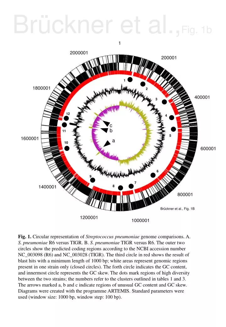
Brückner et al.,Fig. 1b 1 2 3 12 4 c b 11 5 a 10 6 9 7 8 Brückner et al., Fig. 1B Fig. 1. Circular representation of Streptococcus pneumoniae genome comparisons. A. S. pneumoniae R6 versus TIGR. B. S. pneumoniae TIGR versus R6. The outer two circles show the predicted coding regions according to the NCBI accession number NC_003098 (R6) and NC_003028 (TIGR). The third circle in red shows the result of blast hits with a minimum length of 1000 bp; white areas represent genomic regions present in one strain only (closed circles). The forth circle indicates the GC content, and innermost circle represents the GC skew. The dots mark regions of high diversity between the two strains; the numbers refer to the clusters outlined in tables 1 and 3. The arrows marked a, b and c indicate regions of unusual GC content and GC skew. Diagrams were created with the programme ARTEMIS. Standard parameters were used (window size: 1000 bp, window step: 100 bp).
1 2 6 c a 5 4 3 Brückner et al., Fig. 1A Brückner et al.,Fig. 1a
spr0102 - argG 109,691 110,000 112,000 114,000 116,000 118,000 114,000 115,279 120,000 114,000 116,975 122,000 118,000 SP0117 - pspA 124,000 126,000 126,908 spr0121 - pspA Brückner et al., Fig. 2 Fig. 2. The R6 cluster 1 region. The numbers refer to the borders of sequence divergence between S. pneumoniae TIGR and R6 strains. The arrows depict the orientation of the genes as annotated. Regions with >98% sequence identities are connected by lines. The regions of homology are indicated by different shades of gray.
17/2349/456/496 Hu-11/15/9 R6/D39 2303R ATCC D219 1711 4241 2306 670 F1 S1 4 1 1* 2 2* 3 3* 4* 4 5* 6* 5 7* 6 8* 7 8 9* 9 10* 11* 10 Brückner et al., Fig. 4 11 12 12* 13* Brückner et al.,Fig. 4 Fig. 4. Gene clusters of S. pneumoniae TIGR detectable on oligonucleotide microarrays in hybridization with genomic DNA of genetically distinct S. pneumoniae strains. The genes are arranged according to the annotated genome with the replication start on top. Low hybridization signals are indicated by black lines, and only those genes that differ by an intensity ratio smaller than –4 in at least one of the strains shown are marked by black lines. Clusters that are not contained in the R6 strain are marked by arrows on the left side (1-12); other clusters noticeable in other strains are marked on the right (1* - 13*). The strains used for hybridization are described in detail in (Hakenbeck et al. 2001): strain 4 (serotype 4 from Papua), the pair R6/D39, serotype 22 ATCC 49619, serotype 23F D219 from Germany; serotype 23F F1 (France); three representatives of the Hungarian seroytpe 19A multiresistant clone (Hu-11, Hu-15, Hu-9); the two serotype 3 strains 1711 (Switzerland) and 4241 (France); serotype 23F from Finland; four representatives of the multiresistant Spanish 23F clone 17 (South Africa), 2349, 456 and the 19F capsular variant 496 from Spain; and the multiresistant serotype 6B from Spain.