Download
1 / 50
510 likes | 846 Vues
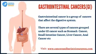
Pictorial lesson in GI Cancers. Oesophageal Cancer. Staging of Oesophageal Cancer. Incidence in USA by pathology. Squamous cell carcinoma. Endoscopic view of the oesophagus shows a tiny, early ulcer which proved on biopsy to be malignant. Squamous cell carcinoma .

E N D
Squamous cell carcinoma • Endoscopic view of the oesophagus shows a tiny, early ulcer which proved on biopsy to be malignant.
Squamous cell carcinoma • An established, infiltrating, well-differentiated lesion exhibits islands of malignant epithelium invading deep into oesophageal muscle.
Squamous cell carcinoma • Endoscopic view shows circumferential involvement of the oesophagus with friable tumour. • Note the narrowed lumen.
CASE: Squamous cell carcinoma • A 62-year-old man with progressive dysphagia and marked weight loss was found on endoscopy to have a poorly differentiated tumour of the middle third of the oesophagus. • Barium swallow film shows narrowing of the oesophagus with mucosal destruction, consistent with oesophageal cancer
Case • CT scan reveals regional metastases and a large primary mass obstructing the oesophagus. • Complete clinical remission was achieved in 3 months after combination chemotherapy plus radiotherapy. • Unfortunately, liver metastases subsequently occurred.
Squamous cell carcinoma • This sagittal section through the larynx, trachea and anterior wall of the Oesophagus obtained at autopsy of a 57-year-old man who presented with a short history of dysphagia. • A barium swallow revealed neoplastic obstruction of the oesophagus; the patient died soon afterward from bronchopneumonia. • A solid, raised, pale tumour (6 x 2 x 2 cm), arising in the oesophagus, has infiltrated the posterior wall of the trachea, forming a nodular projection into the tracheal lumen.
Case • Adenocarcinoma. Weight loss and right upper abdominal pain, with minimal dysphagia, developed in a 58-year-old man. • Upper GI endoscopy showed a constricting, poorly differentiated lesion of the lower third of the oesophagus. Barium swallow film defines the extent of the lesion (arrows). • On CT scan, a large liver metastasis
Treatment • Depends on stage, but includes surgery, radiotherapy, chemotherapy and laser treatment
Gastric Cancer • Barium swallow study shows a large fundal carcinoma
Malignant gastric ulcer • This antral lesion exhibits heaped-up nodular margins, particularly suggestive of malignancy
Adenocarcinoma. • This intestinal-type tumour exhibits well-formed malignant glandular elements
Adenocarcinoma • Barium swallow study reveals a large polypoid lesion in the body of the stomach, causing a filling defect.
Diffuse adenocarcinoma - linitis plastica • Barium study shows the typical appearance of an extensive linitis plastica involving the entire stomach, which appears fixed and narrowed. • No peristalsis was observed and barium flowed out of the stomach quickly. • The mucosal edge is only slightly irregular; ulceration of the mucosa may be minimal or absent in this type of carcinoma. • (Arrow indicates the gastric fundus.)
Case • Diffuse adenocarcinoma (linitis plastica). • This gastrectomy specimen, opened anteriorly, is from a 64-year-old man who had a 3-year history of dyspepsia. Barium swallow and endoscopy revealed a gastric carcinoma. • There is diffuse infiltration of the pylorus and body of the stomach by pale tumour, as well as marked luminal narrowing, although the tumour has no exophytic component. • Note the irregular infiltration of the muscle coat.
Pancreatic carcinoma • Barium study shows a tumour mass in the head of the pancreas invading the duodenal loop and producing changes in the fold pattern.
Pancreatic carcinoma • Abdominal CT scan shows a large focal mass in the tail of the pancreas
Staging of hepatocellular carcinoma (including intrahepatic bile ducts)
Multifocal hepatocellular carcinoma in haemochromatosis • A 61-year-old man with haemochromatosis and a 12-year history of hepatomegaly and diabetes mellitus died after developing liver failure with ascites. • Section through the right lobe of the liver shows an ill-defined micronodular cirrhosis, associated with deep brick-red parenchymal pigmentation. • Scattered throughout the posterior region are many pale nodules of carcinoma.
Primary hepatic angiosarcoma • Typically, these tumours may appear as (a) a surface vascular tumour or as a hemorrhagic tumour mass. • They are associated with industrial exposure to vinyl chloride and the radiographic contrast agent Thorotrast and usually comprise multicentric hemorrhagic nodules.
Lung metastases • Chest film of a 19-year-old Asian man who presented with hepatocellular carcinoma shows the well-defined pulmonary nodules characteristic of metastatic deposits. Rapid disease progression occurred within 2 months. • Metastases are unusual with hepatoma but do occur to the bones, lung and brain.
Familial adenomatous polyposis • FAP is a genetic disorder greatly increasing the risk of bowel cancer. • Barium enema study demonstrates multiple, small polyps throughout the colon.
Familial adenomatous polyposis • Innumerable adenomatous polyps, increasing in size and density from proximal (upper left) to distal (lower right)
Carcinoma in ulcerative colitis • Malignancies developing in ulcerative colitis may present as an infiltrative plaque, or a polypoid mass • The cumulative risk of cancer increases dramatically with the duration of ulcerative colitis. After 20 years, there is a 15% incidence of colon cancer, which increases to 50% after 40 years.
Carcinoma in ulcerative colitis • They may also develop as a stricture.
Modified Dukes' staging classification of colorectal cancer.
Staging • Stages B3 and C3 (not shown) signify perforation or invasion of contiguous organs or structures (T4). • The TNM classification provides a more accurate staging system: Dukes B is a composite of better (T2N0) and worse (T3N0, T4N0) prognostic groups as is Dukes C (TxN1 or TxN2).
Adenocarcinoma of caecum • Intestinal obstruction occurs late in the course of the disease. • Although this lesion (arrows) is relatively large, there was no obstruction to retrograde filling of the ileum and no dilatation of the small intestine. • Symptoms may include anaemia or dyspepsia and weight loss reminiscent of a benign or malignant gastric ulcer.
Adenocarcinoma of caecum • Large, fungating tumours, as seen here, are a less common presentation of colorectal tumours; they predominate in the caecum.
Adenocarcinoma • Moderately differentiated tumours are marked by gland (acinar) formation by malignant epithelium; there is considerable nuclear pleomorphism within individual cells
Adenocarcinoma of sigmoid colon • Barium enema shows an annular stenosing lesion of the distal sigmoid, producing a characteristic 'apple core' appearance
Adenocarcinoma of colon. • This specimen exhibits an annular, stenosing lesion with dilatation of the colon proximal to it. • This appearance may be seen at any site and is facilitated by circumferential spread of the tumour through submucosal (or serosal) lymphatic channels
Treatment • Surgical, even if palliative to relieve obstruction • Adjuvant chemotherapy if node positive or at increased risk of metastases
Adenocarcinoma of rectum • This lower rectal lesion demonstrates the most common macroscopic appearance of colorectal cancers as well-circumscribed lesions with raised edges and an ulcerated centre.
Treatment • Combination of surgery, radiotherapy and chemotherapy, depending on extent of disease
Anatomy of the lower rectum and anal canal. • The anal canal extends from the anorectal ring to an area about halfway between the dentate (pectinate) line and the anal verge. • The anal margin consists of the area distal to the anal canal, including the perianal skin
Squamous cell carcinoma of anal margin • Squamous cancers of the anus are divided into tumours arising in the anal canal (most often above the dentate line) and those arising in the skin at the anal margin, as shown here. • This lesion measures 1cm across. Neoplasms at this site tend to be slow growing and metastasize to inguinal lymph nodes. • They have a 5-year survival rate of approximately 70%.
Anal Cancer • Treatment is potentially curative with combined chemo radiotherapy • These tumours are often HPV related and associated with immuno-supression.
Malignant melanoma of anal canal • This specimen is from a 74-year-old woman who presented with a brief history of episodic rectal bleeding. • A hard mass was palpable in the lateral wall of the anal canal and an abdominoperineal resection was performed. • The anal canal has been opened to show a flattened, ovoid nodule (2 cm in diameter) arising at about the level of the dentate line. The edge of the tumour shows obvious melanotic pigmentation and an irregular streak of pigment extends from the nodule to the anal margin. • Anorectal melanoma is rare, accounting for about 1% of anal cancers.