Download
1 / 17
170 likes | 364 Vues
March 22, 2010 By: Catherine Roberts. EPITHELOID SARCOMA LEFT THIGH. History:.

E N D
March 22, 2010 By: Catherine Roberts EPITHELOID SARCOMA LEFT THIGH
History: This 43-year-old, right-handed male presents today with a lump in his left thigh. He states that he first noticed this lump in June 2003. He noticed that it hurt when things in his pocket, for example his keys, hit it. He thought it would go away and waited the entire summer before seeking treatment. He saw his primary care physician in October 2003. He performed an MRI and then referred the patient to an orthopedic surgeon. The orthopedic surgeon reviewed the MRI and felt that this was likely cancerous and then referred him here to Mayo. Mr. Marshall states that since June of this year he feels that his lump has been getting a little bit bigger and is very, very slightly painful. He is not taking any medicines for the pain. He states that it does not really interfere with ambulation. It also becomes somewhat painful if he sits for a long period of time.
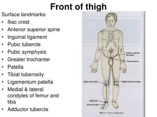
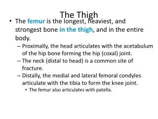
Findings: 2/11/04 MRI - There is a large mass confined to the left vastus medialis that abuts the anterolateral periosteum of the femur without cortical or intraosseous invasion. The mass is somewhat heterogeneous and contains at least one fluid-filled area, probably representing necrosis anteriorly. The mass measures approximately 8.2 x 3.6 x 5.4 cm in the superoinferior, anteroposterior, and mediolateral dimensions respectively. There is no involvement of the neurovascular bundle. 6/24/04 MRI s/p chemotherapy and radiation - The known epithelioid sarcoma located within the vastus intermedius muscle of the anterior left midthigh has significantly decreased in size from 02/11/04. The tumor now measures 2.7 x 4.7 x 6.7 cm. The borders of the mass have become less convex and the tumor mass has less intense enhancement with scattered regions of nonenhancing central necrosis. 6/30/04 - Radical excision soft tissue sarcoma; insertion of brachytherapy tubes.
Findings (Continued): Surgical resection followed by placement of after-loading tubes for perioperative brachytherapy.
Diagnosis: Excision of left thigh mass: Malignant tumor with features of high-grade proximal type epithelioid sarcoma forming a mass 8 x 5 x 4.1 cm with some fibrosis and cytologic changes consistent with prior chemoradiation. The specimen includes skin with scar consistent with prior biopsy site.
General Discussion: Epithelioid sarcoma. Accounts for 1.7% of all sarcomas. Approximately 170 new cases per decade recorded at AFIP. Predilection for upper extremities, especially the hand and wrist.
References: Kransdorf and Murphey, Imaging of Soft Tissue Tumors. WB Sanders 1997, Pages 361-367.