Download
1 / 19
190 likes | 203 Vues
Explore the functions of cellular membranes in controlling the movement of ions and electrical signaling. Discover how voltage-gated channels, transporters, and receptors play a role in transmitting signals across nerve cells.

E N D
FIGURE 14.1. The positively charged potassium ion cannot cross the lipid bilayer, but passes easily through a water-filled tube in a potassium channel.
FIGURE 14.2. The resting voltage of (A) glial and (B) nerve cells.
FIGURE 14.3. A nerve cell from the retina viewed with its nutritive capillaries. Red blood cells are an example of cells whose membrane voltage varies little, while nerve cells are specialized to transmit electrical signals over long distances. Image by Professor David Becker, University College London; used with permission.
FIGURE 14.4. Chloride is close to equilibrium across most plasma membranes.
FIGURE 14.5. The glucose carrier and the sodium/calcium exchanger are sometimes open to the cytosol and sometimes open to the extracellular medium.
FIGURE 14.7. The calcium ATPase undergoes a cycle of phosphorylation and dephosphorylation. These drive shape changes that in turn push calcium ions out of the cell.
FIGURE 14.10. One type of pain receptor has a distal terminal in the skin and is connected to the central nervous system by a myelinated nerve fiber.
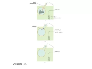
FIGURE 14.11. Electrical events in a pain receptor nerve cell.