Download
1 / 34
340 likes | 560 Vues
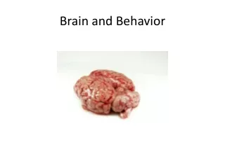
Chapter 2 Brain and Behavior. Neuron and Its Parts. Neuron: Individual nerve cell Dendrites: Receive messages from other neurons Soma: Cell body; body of the neuron Axon: Fiber that carries information away from the cell body

E N D
Neuron and Its Parts • Neuron: Individual nerve cell • Dendrites: Receive messages from other neurons • Soma: Cell body; body of the neuron • Axon: Fiber that carries information away from the cell body • Axon Terminals: Branches that link the dendrites and somas of other neurons
Figure 2.1 FIGURE 2.1 A neuron, or nerve cell. In the right foreground you can see a nerve cell fiber in cross section. The upper left photo gives a more realistic picture of the shape of neurons. Nerve impulses usually travel from the dendrites and soma to the branching ends of the axon. The nerve cell shown here is a motor neuron. The axons of motor neuron stretch from the brain and spinal cord to muscles or glands of the body.
Synapses • Messages from one neuron to another pass over a microscopic gap called a synapse • Synapse: Microscopic gap between two neurons over which messages pass
Figure 2.5 FIGURE 2.5 A highly magnified view of a synapse. Neurotransmitters are stored in tiny sacs called synaptic vesicles (VES-ihkels). When a nerve impulse reaches the end of an axon, the vesicles move to the surface and release neurotransmitters. These molecules cross the synaptic gap to affect the next neuron. The size of the gap is exaggerated here; it is actually only about one millionth of an inch. Some transmitter molecules excite the next neuron, and some inhibit its activity.
Neurotransmitters • Chemicals that alter activity in neurons; brain chemicals • Acetylcholine: Activates muscles • Dopamine: Muscle control • Serotonin: Mood and appetite control • Receptor Site: Areas on the surface of neurons and other cells that are sensitive to neurotransmitters
Nerves and Neurons • Nerves: Large bundles of axons and dendrites • Myelin: Fatty layer of tissue that coats axons • Multiple Sclerosis (MS) occurs when myelin layer is destroyed; numbness, weakness, and paralysis occur
Neural Networks • Central Nervous System (CNS): Brain and spinal cord • Peripheral Nervous System: All parts of the nervous system outside of the brain and spinal cord • Somatic System: Links spinal cord with body and sense organs; controls voluntary behavior • Autonomic System: Serves internal organs and glands; controls automatic functions such as heart rate and blood pressure
Figure 2.6 FIGURE 2.6 (a) Central and peripheral nervous systems. (b) Spinal nerves, cranial nerves, and the autonomic nervous system.
Two Divisions of the Autonomic System • Sympathetic: Arouses body; emergency system • Parasympathetic: Quiets body; most active after an emotional event
Figure 2.8 FIGURE 2.8 Sympathetic and parasympathetic branches of the autonomic nervous system. Both branches control involuntary actions. The sympathetic system generally activates the body. The parasympathetic system generally quiets it. The sympathetic branch relays its messages through clusters of nerve cells outside the spinal cord.
The Spinal Cord • Spinal Nerves: 31 of them; carry sensory and motor messages to and from the spinal cord • Cranial Nerves: 12 pairs that leave the brain directly; also work to communicate messages
Figure 2.7 FIGURE 2.7 Subparts of the nervous system.
Figure 2.9 FIGURE 2.9 A sensory-motor arc, or re- flex, is set in motion by a stimulus to the skin (or other part of the body). The nerve impulse travels to the spinal cord and then back out to a muscle, which contracts. Such reflexes provide an “automatic” protective device for the body.
Researching the Brain • Ablation: Surgical removal of parts of the brain. • Deep Lesioning: A thin wire electrode is lowered into a specific area inside the brain. Electrical current is then used to destroy a small amount of brain tissue. • Electrical Stimulation of the Brain (ESB): When an electrode is used to activate target areas in the brain. • Electroencephalograph (EEG): Detects, amplifies, and records electrical activity in the brain.
Figure 2.10 FIGURE 2.10 The functions of brain structures are explored by selectively activating or removing them. Brain research is often based on electrical stimulation, but chemical stimulation is also used at times.
Researching the Brain (cont'd) • Computed Tomographic Scanning (CT): Computer-enhanced X-ray image of the brain or body • Magnetic Resonance Imaging (MRI): Uses a strong magnetic field, not an X-ray, to produce an image • Functional MRI (fMRI): MRI that also records brain activity • Positron Emission Tomography (PET): Computer-generated color image of brain activity, based on glucose consumption in the brain
Cerebral Cortex • Definition: Outer layer of the cerebrum • Cerebrum: Two large hemispheres that cover upper part of the brain • Corticalization: Increase in size and wrinkling of the cortex • Cerebral Hemispheres: Right and left halves of the cortex • Corpus Callosum: Bundle of fibers connecting cerebral hemispheres
Figure 2.21 FIGURE 2.21 The left and right brain have different information-processing styles. The left brain focuses on the small details; the right gets the big pattern.
Split Brains • Corpus Callosum is cut; done to control severe epilepsy (seizure disorder). • Result: The person now has two brains in one body. • This operation is rare and is often used as a last resort.
Figure 2.19 FIGURE 2.19 Basic nerve pathways of vision. Notice that the left portion of each eye connects only to the left half of the brain; likewise, the right portion of each eye connects to the right brain. When the corpus callosum is cut, a “split brain” results. Then visual information can be sent to just one hemisphere by flashing it in the right or left visual field as the person stares straight ahead.
Figure 2.20 FIGURE 2.20 A circle is flashed to the left brain of a split-brain patient, and he is asked what he saw. He easily replies, “A circle.” He can also pick out the circle by merely touching shapes with his right hand, out of sight behind a screen. However, his left hand can’t identify the circle. If a triangle is flashed to the patient’s right brain, he can’t say what he saw (speech is controlled by the left hemisphere). He also can’t identify the triangle by touch with the right hand. Now, however, the left hand has no diffi- culty picking out the triangle. In other tests, the hemispheres reveal distinct skills, as listed above the drawing.
Central Cortex Lobes • Occipital: Back of brain; vision center • Parietal: Just above occipital; bodily sensations such as touch, pain, and temperature • Temporal: Each side of the brain; auditory and language centers • Frontal: Movement, sense of smell, higher mental functions • Contains motor cortex; controls motor movement
Figure 2.23 FIGURE 2.23 The lobes of the cerebral cortex and the primary sensory, motor, and association areas on each. The top diagrams show (in cross section) the relative amounts of cortex “assigned” to the sensory and motor control of various parts of the body. (Each cross section, or “slice,” of the cortex has been turned 90 degrees so you see it as it would appear from the back of the brain.)
Figure 2.25 FIGURE 2.25 This simplified drawing shows the main structures of the human brain and describes some of their most important features. (You can use the color code in the foreground to identify which areas are part of the forebrain, midbrain, and hindbrain.)
Forebrain • Structures are part of Limbic System: System within forebrain closely linked to emotional response • Thalamus: Relays sensory information to the cortex; switchboard • Hypothalamus: Regulates emotional behaviors and motives (e.g., sex, hunger, rage, hormone release) • Hippocampus: Associated with storing memories;
Endocrine System • Glands that pour chemicals (hormones) directly into the bloodstream or lymph system • Pituitary Gland: Regulates growth via growth hormone • Too little means person will be smaller than average • Hypopituitary Dwarfs: As adults, perfectly proportioned but tiny • Too much leads to giantism • Excessive body growth
Endocrine System (cont'd) • Acromegaly: Enlargement of arms, hands, feet, and facial bones • Too much growth hormone released late in growth period • Andre the Giant
Endocrine System Concluded • Pineal Gland: Regulates body rhythms and sleep cycles. • Releases hormone melatonin, which responds to daily variations in light. • Thyroid: In neck; regulates metabolism. • Hyperthyroidism: Overactive thyroid; person tends to be thin, tense, excitable, nervous. • Hypothyroidism: Underactive thyroid; person tends to be inactive, sleepy, slow, obese.
The Adrenal Glands • Adrenals: Arouse body, regulate salt balance, adjust body to stress, regulate sexual functioning; located on top of kidneys • Releases epinephrine and norepinephrine (also known as adrenaline and noradrenaline) • Epinephrine arouses body; is associated with fear • Norepinephrine arouses body; is linked with anger