Download
1 / 43
430 likes | 651 Vues
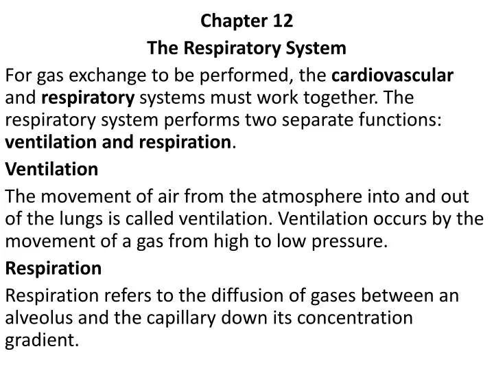
Chapter 12 The Respiratory System For gas exchange to be performed, the cardiovascular and respiratory systems must work together. The respiratory system performs two separate functions: ventilation and respiration . Ventilation

E N D
Chapter 12 The Respiratory System For gas exchange to be performed, the cardiovascular and respiratory systems must work together. The respiratory system performs two separate functions: ventilation and respiration. Ventilation The movement of air from the atmosphere into and out of the lungs is called ventilation. Ventilation occurs by the movement of a gas from high to low pressure. Respiration Respiration refers to the diffusion of gases between an alveolus and the capillary down its concentration gradient.
PathophysiologicConcepts Atelectasis The collapse of either a lung or an alveolus is called atelectasis. This results in a reduction in the surface area available for diffusion. Newborns may be born with alveoli collapsed at birth. This condition is called primary atelectasis. The collapse of previously expanded alveoli is called secondary atelectasis. The two main types of atelectasis are : a- Compression Atelectasis This occurs if the chest wall is punctured or opened because atmospheric pressure is greater than the pressure holding the lungs expanded (pleural pressure). Compression atelectasis can also occur from a growing tumor or abdominal distention.
b-Absorption Atelectasis Any situation that results in mucus accumulation, such as cystic fibrosis, pneumonia, or chronic bronchitis, anesthesia and prolonged bed rest after surgery may lead to absorption atelectasis. Mucus accumulation increases the risk of pneumonia because mucus can act as a breeding ground for growth of microorganisms. Hypoxemia Reduced oxygen concentration in arterial blood is called hypoxemia. Hypoxemia can occur if there is decreased oxygen in the air (hypoxia) or atelectasis.
Pulmonary Hypertension Elevated blood pressure in the pulmonary vascular system is called pulmonary hypertension. Causes of Pulmonary Hypertension (1) a prolonged increase in pulmonary blood flow (2) an increase in pulmonary resistance to flow . Consequences of Pulmonary Hypertension -A right-sided heart failure called corpulmonale - Pulmonary edema because the capillary hydrostatic force favoring filtration is increased.
Conditions of Disease or Injury Upper Respiratory Tract Infections (URTI) Infections caused by any microorganism of the nongas-exchanging upper structures (the nasal passages, the pharynx, and the larynx). It includes the common cold, pharyngitis or sore throat, laryngitis, and uncomplicated influenza. usually caused by viruses, although bacteria may also be involved either initially or secondary to a viral infection. The inflammatory reaction leads to increased mucus production
Clinical Manifestation - Cough - Sneezing and nasal congestion - Mucus production - Headache - Low-grade fever - Malaise (physical discomfort) Diagnostic Tools A good history and physical examination will assist diagnosis. Complications - Sinusitis and acute otitis media may develop. - Lower respiratory tract infections, including pneumonia and bronchitis.
Treatment - Rest to reduce the body's metabolic demands. - Extra hydration helps liquefy the thick mucus, making it easier to move it out . - Decongestants, antihistamines, and cough suppressants may provide some symptom relief. - Antibiotics are required if the infection is bacterial . Notice: Effects of Cigarette Smoking on Respiratory Defenses • Stimulate mucus production • Paralyze the cilia. • increasing the risk of microbial growth.
Pneumonia Pneumonia, an acute infection of the lung tissue are bacterial in origin, occurring as a primary condition or secondary to a previous viral infection. The most common cause is Streptococcus pneumoniae. Clinical Manifestations - Fever and chills and a cough that is often productive, purulent, and present throughout the day; infants may grunt in an attempt to improve airflow. - Sputum that is rust colored (for Streptococcus pneumoniae), pink (for Staphylococcus aureus), or greenish with a particular odor (for Pseudomonas aeruginosa).
- Wheezing, signifies obstruction to airflow. - Significantly increased respiratory rate. - Fatigue, from both inflammatory reactions and hypoxia. - Pleural pain from inflammation and edema. - Dyspnea - Hemoptysis, the coughing up of blood. - Chest pain as a result of pleural irritation.
Diagnostic Tools - White blood cell count generally increases . - Edema of the interstitial space is often apparent on chest radiograph (x-ray). - Arterial blood gases may be abnormal. Complications - Cyanosis with accompanying hypoxia may develop. - Absorption atelectasis. - Respiratory failure and death. Treatment - Pretreatment sputum sample determines the treatment . - Antibiotics - Rest. - Hydration to help loosen secretions.
Tuberculosis It is caused by the microorganism Mycobacterium tuberculosis.It can enter by inhalation or by means of ingestion of contaminated unpasteurized milk. Risk Factors for Tuberculosis : - living in close quarters with someone who has an active infection. - workers caring for the infected individuals - individuals who have inadequate immune systems.
Clinical Manifestations If active tuberculosis develops, an individual usually demonstrates the following: - Fevers, especially in the afternoon. - Malaise. - Night sweats. - Loss of appetite and weight loss. - A productive, purulent cough accompanied by chest pain is common with active infection.
Diagnostic Tools - A positive skin test for tuberculosis . - A sputum sample followed by microscopic examination or culturing - Chest radiograph demonstrates current or previous tubercle formation. Complications Severe disease may lead to overwhelming sepsis, respiratory failure, and death. Treatment Currently, treatment of individuals who have an active infection includes a combination of four drugs and lasts at least 9 months or longer.
Pneumothorax Pneumothorax is the collapse of all or part of a lung that occurs when air or another gas enters the pleural space surrounding the lungs. There are two different types of pneumothorax: Open and Spontaneous Pneumothorax -An open pneumothorax occurs when the chest wall has been opened and air is allowed into the pleural space from the atmosphere leading to collapses. -A spontaneous pneumothorax occurs when the chest wall is intact, but the lungs begin to leak air into the pleural space .
Tension Pneumothorax In this case, air enters the pleural space during inspiration and cannot move back into the lungs with expiration because the small hole collapses as the lungs deflate. Atelectasis and displacement of the heart and great vessels may result in severe alterations of cardiovascular function.
Clinical Manifestations - Acute onset of pain . - Rapid, shallow breathing (tachypnea) and dyspnea are common. - The chest appears asymmetrical. Tracheal deviation may also be apparent. Diagnostic Tools - Blood gases and hemoglobin saturation will indicate hypoxia. - Radiographs can identify a collapsed lung. Complications - A tension pneumothorax may lead to reduced cardiac filling . • Hypoxia and severe dyspnea. • Death may occur.
Treatment - A tension pneumothorax is treated immediately with insertion of a chest tube or a large-bore needle into the pleural space with subsequent suction of the air out of the space. - A small spontaneous pneumothorax is treated by insertion of a chest tube connected to a drainage tube that is kept in place until the pleural injury is healed. - Any penetrating wound should be covered or closed.
Respiratory Failure Inadequate exchange of gas that results in hypoxia, hypercapnia (increased arterial carbon dioxide concentration), and acidosis is called respiratory failure Clinical Manifestations - Cyanosis. - Severe dyspnea. Diagnostic Tools -partial pressure of oxygen in arterial blood of less than 50 mmHg, and a partial pressure of carbon dioxide in arterial blood of greater than 50 mmHg, with a pH less than or equal to 7.25.
Complications - Multi-organ failure. - Death. Treatment Oxygen support, including artificial ventilation, is required. In general, the sooner a person is put on ventilatory support, the better the prognosis.
Respiratory Distress Syndrome of the Newborn (hyaline membrane disease) Is a condition of pulmonary hypoxia resulting from widespread primary atelectasis. Hypoxia develops, leading to pulmonary injury and a subsequent inflammatory reaction with the accumulation of white blood cells and production of hyaline membranes, which are white fibrin accumulations lining the alveoli.
Risk Factors for Respiratory Distress Syndrome - The primary risk factor is prematurity. 5 and 10% suffer from this syndrome. The more premature the infant, the more likely RDS will develop. The alveolar cells that produce surfactant do not mature until between 28 and 32 weeks of gestation. Alveoli of premature infants are small and their chest muscles are weak. - Infants born to insulin-dependent diabetic mothers.
Clinical Manifestations - Increased respiratory rate. - Intercostal or chest retractions with each breath. - Nasal flaring with each breath. Diagnostic Tools - The clinical appearance coupled with the pregnancy history. - Arterial blood gases . - Chest radiograph typically shows diffuse granular densities within hours of birth.
Complications - Some infants who survive RDS develop a chronic respiratory disease characterized by alveolar scarring, inflammation of the alveoli and the capillaries, and pulmonary hypertension. - Signs of dyspnea and hypoxia may continue and proceed to infant exhaustion, respiratory failure, and death, usually within 3 days.
Treatment - Delay of parturition (delivery of an infant) for even 24 to 48 hours has been shown to reduce the incidence and severity of RDS. This is because the stress of labor increases maternal and fetal cortisol release from the adrenal cortex which stimulate alveolar cells to produce surfactant. - Maternal injections of corticosteroids at least 24 hours before a premature infant is delivered can significantly reduce the incidence of RDS. -
-Treatment is supportive and consists of oxygen therapy, maintenance of a quiet, warm environment to decrease oxygen requirements, nutritional support, and repeated evaluation of blood gases and acid-base status. - A major treatment advance has been the development of artificial surfactant delivered directly into the lower respiratory tract This treatment, combined with maternal corticosteroid injections, offers the best hope for reducing the morbidity and mortality caused by RDS. - Mechanical ventilation may be employed to treat RDS.
Sudden Infant Death Syndrome Characterized by the unexpected and the unexplained death of a previously healthy infant, between 1 week and 1 year of age. The highest incidence of SIDS is between 2 and 4 months of age, and occurs primarily during the night. Causes of Sudden Infant Death Syndrome The cause of SIDS is unknown. Some evidence suggests that an immature central nervous system fails to respond appropriately to increasing levels of carbon dioxide.
Cystic Fibrosis Cystic fibrosis is a hereditary disease characterized by alterations of exocrine gland function throughout the body. It results in production of large amounts of thick mucus and increased concentration of sodium and chloride in the sweat. Effects of Cystic Fibrosis The main body systems affected by the mucus accumulation are the pulmonary and the gastrointestinal systems. Other organs also are victims of the excess mucus, including the liver and the reproductive organs. Sweat glands over-secrete sodium chloride, and sweat accumulates on the skin.
Clinical Manifestations -A protuberant abdomen may be apparent soon after birth, resulting from an inability to pass meconium in the first stool. -Salty taste when kissed, caused by salt buildup from sweat on the skin. -Repeated respiratory tract infections throughout infancy and early childhood. -Chronic rhinitis, and chronic cough and sputum production. -Failure to thrive because nutrients are poorly absorbed.
Asthma Asthma is a progressive respiratory disease characterized by inflammation of the respiratory tract and spasm of bronchiolar smooth muscle. This results in excess mucus production and a decrease in ventilation of the alveoli. Risk factors include: - a family history of asthma or allergy. - repeated upper respiratory infections may also trigger adult-onset asthma. - occupational exposure to dusts and irritants.
Clinical Manifestations -Significant dyspnea. -Coughing, especially at night. -Rapid, shallow breathing. -Audible wheezing heard only on expiration. -Chest retractions and, with a worsening of condition, nasal flaring. -Anxiety, related to the inability to get enough air.
Complications - Status asthmaticus, a life-threatening condition of prolonged bronchiolar spasm that cannot be reversed with medication. Treatment - For all stages of asthma, prevention of exposure to known allergens is vital. - Oral or inhaled corticosteroids early in the course of an attack or as preventive therapy. - Bronchodilators
Acute Bronchitis Bronchitis is a common, obstructive respiratory disease consisting of inflammation of the bronchi. It is usually associated with a viral or a bacterial infection or the inhalation of irritants such as cigarette smoke or chemicals present in air pollution. It is characterized by excess mucus production. Clinical Manifestations - Cough, usually productive with thick mucus and purulent sputum. - Dyspnea. - Fever. - Hoarseness. - Crackles (discontinuous fine or coarse lung sounds), especially on inspiration. - Chest pain occasionally may be present.
Diagnostic Tools -Chest radiograph. Complications -Repeated episodes of acute bronchitis may result in the pathologic changes characteristic of chronic bronchitis. Treatment - Antibiotics for secondary or primary bacterial infections. - Increased fluid intake and expectorants to loosen mucus. - Rest to reduce oxygen demands.
Chronic Bronchitis It is an obstructive pulmonary disorder characterized by excessive mucus production in the lower respiratory tract and a resulting chronic cough. It must last for at least 3 consecutive months of the year for 2 consecutive years. Clinical Manifestations - A productive, purulent cough, worsened by inspired irritants, cold weather, or an infection. - Copious amounts of sputum production. - Air hunger and dyspnea.
Diagnostic Tools - Pulmonary function tests demonstrate a reduction in vital capacity. - Blood gases show decreased arterial oxygen and increased arterial carbon dioxide. - Chest radiograph may document chronic bronchitis and fibrosis of the lung tissue.
Complications - Pulmonary hypertension. - Clubbing of the end segment of the fingers - Polycythemia , hypoxia stimulate erythropoietin secretion. - Lung cancer. Treatment - Decreasing further irritant exposure, especially cigarette smoke. - Prophylactic antibiotic therapy. - Bronchodilators are frequently prescribed. - Anti-inflammatory drugs reduce mucus production and relieve blockage. - Expectorants and increased fluid intake loosen the mucus. - Oxygen therapy may be required.
Emphysema Is a chronic obstructive disease characterized by loss of lung elasticity and a reduction in alveolar surface area due the destruction of the alveolar walls and the enlargement of air spaces distal to the terminal bronchioles. Clinical Manifestations - Air trapping, resulting from the loss of lung elasticity and leading to expansion of the chest (increased anterior-posterior diameter). - Diminished breath sounds on auscultation. - Use of accessory muscles of respiration. - Tachypnea (increased respiratory rate) caused by hypoxia and hypercapnia. - Central nervous system depression, resulting from high carbon dioxide levels .
N.B.One key difference between emphysema and chronic bronchitis is the lack of sputum production in emphysema. Diagnostic Tools • Abnormal pulmonary function tests, decreased vital capacity, and increased residual volume - As the disease progresses, blood-gas analysis will first demonstrate hypoxia. Late in the disease, carbon dioxide levels may also be elevated. Complications - Pulmonary hypertension . - A reduction in quality of life is common in severely affected individuals.
Treatment -There is no cure . -Relieving the symptoms and preventing a worsening of the condition is the objective in emphysema treatment. - Encouraging the individual to stop smoking. - Oxygen therapy - Well-designed exercise therapy can improve symptoms.
Chronic Obstructive Pulmonary Disease Individuals who have long-standing emphysema also usually have chronic bronchitis and demonstrate indications of both diseases. This condition is called chronic obstructive pulmonary disease (COPD). Clinical Manifestations -Symptoms of both emphysema and chronic bronchitis are usually present. - Dyspnea is constant. Diagnostic Tools - History and physical examination. - Chest x-ray.
Complications - Pulmonary hypertension leading to corpulmonale. - Pneumothorax. Treatment In general, COPD treatment is as described for chronic bronchitis and emphysema.
Lung Cancer Lung cancer is defined as a cancer of the epithelial lining of the respiratory tract (bronchogenic carcinoma). It can occur anywhere in the lung. Risk Factors for Lung Cancer - Cigarette smoking - Chronic bronchitis Clinical Manifestations - A persistent cough. - Recurring lower respiratory tract infections. - Hemoptysis (the coughing up of blood). -Weight loss. - Fatigue. - Hoarseness. - Pain or dysfunction in a distant organ reflecting metastasis may be the first clue of lung cancer.
Diagnostic Tools Chest x-ray followed by biopsy of suspicious lesions may diagnose the disease. Complications Prognosis is poor. The 5-year survival rate for all types of lung cancer is only 13%. Some types of lung cancer have an even worse prognosis. Treatment Any combination of surgery, radiation, and chemotherapy.