Download
1 / 119
1.2k likes | 1.4k Vues
Unit 7 – The Nervous System. Contains 2 kinds of cells: neurons: cells that send and receive signals neuroglia ( glial cells): cells that support and protect neurons. Neural Tissue. Brain and spinal cord Sensory receptors of sense organs (eyes, ears, etc.)

E N D
Contains 2 kinds of cells: • neurons: • cells that send and receive signals • neuroglia (glial cells): • cells that support and protect neurons Neural Tissue
Brain and spinal cord Sensory receptors of sense organs (eyes, ears, etc.) Nerves connect nervous system with other systems Organs of the Nervous System
Central nervous system (CNS) Peripheral nervous system (PNS) Anatomical Divisions of the Nervous System
Consists of the spinal cord and brain Contain neural tissue, connective tissues, and blood vessels The Central Nervous System (CNS)
Are to process and coordinate: • sensory data: • motor commands: • higher functions of brain: • intelligence, memory, learning, emotion Functions of the CNS
Ranges from 750 cc to 2100 cc Contains almost 98% of the body’s neural tissue Average weight about 1.4 kg (3 lb) The Human Brain
There are 3 layers (meninges) that surround the brain and spinal cord • Pia mater – covers the brain • Arachnoid – middle layer • Dura mater – outermost layer that is attached to the interior of the skull Meninges
Cerebrum Cerebellum Diencephalon Mesencephalon Pons Medulla oblongata 6 Regions of the Brain
Largest part of brain Controls higher mental functions Memory storage Divided into left and right cerebral hemispheres Surface layer of gray matter (neural cortex) Cerebrum
Also called cerebral cortex Folded surface increases surface area Elevated ridges (gyri) Shallow depressions (sulci) Deep grooves (fissures) Neural Cortex
Second largest part of brain Coordinates repetitive body movements 2 hemispheres Covered with cerebellar cortex Cerebellum
Located under cerebrum and cerebellum Links cerebrum with brain stem Contains thalamus and hypothalamus Diencephalon
Thalamus: • relays and processes sensory information • Hypothalamus: • hormone production • emotion • thirst/hunger • body temp. • controls circadian rhythms (day–night cycles) Thalamus and Hypothalamus
Processes infomation between: • spinal cord and cerebrum or cerebellum • Includes: • mesencephalon • pons • medulla oblongata The Brain Stem
Also called midbrain Processes sight, sound, and associated reflexes Maintains consciousness Mesencephalon
Connects cerebellum to brain stem Is involved in somatic and visceral motor control Pons
Connects brain to spinal cord • Relays information • Regulates autonomic functions: • Cardiovascular, respiratory, and digestive systems Medulla Oblongata
Is the largest part of the brain Controls all conscious thoughts and intellectual functions Processes somatic sensory and motor information The Cerebrum
The Cerebral Cortex Figure 14–12b
Gyri of neural cortex: • increase surface area (number of cortical neurons) Structures of the Cerebrum (1 of 3)
Longitudinal fissure: • separates cerebral hemispheres • Lobes: • divisions of hemispheres Structures of the Cerebrum (2 of 3)
Central sulcus divides: • anterior frontal lobe from posterior parietal lobe • Lateral sulcus divides: • frontal lobe from temporal lobe • Corpus Callosum • Connects left and right hemispheres Structures of the Cerebrum (3 of 3)
Each cerebral hemisphere receives sensory information from, and sends motor commands to, the opposite side of body The 2 hemispheres have different functions although their structures are alike Correspondence between a specific function and a specific region of cerebral cortex is not precise 3 Functional Principles of the Cerebrum
Central sulcus separates motor and sensory areas Motor and Sensory Areas of the Cortex Figure 14–15a
Precentral gyrus of frontal lobe: • directs voluntary movements Motor Areas
Postcentral gyrus of parietal lobe: • receives somatic sensory information (touch, pressure, pain, vibration, taste, and temperature) Sensory Areas
Visual cortex: • information from sight receptors • Auditory cortex: • information from sound receptors • Olfactory cortex: • information from odor receptors • Gustatory cortex: • information from taste receptors Special Sensory Cortexes
Sensory association areas: • monitor and interpret arriving information at sensory areas of cortex • Somatic motor association area (premotor cortex): • coordinates motor responses (learned movements) Association Areas
Somatic sensory association area: • interprets input to primary sensory cortex (e.g., recognizes and responds to touch) • Visual association area: • interprets activity in visual cortex • Auditory association area: • monitors auditory cortex Sensory Association Areas
Also called Wernicke’s area Present in only 1 hemisphere (left) Receives information from all sensory association areas Coordinates access to complex visual and auditory memories General Interpretive Area Wernicke’s area
Speech center (Broca’s Area): • is associated with general interpretive area • coordinates all vocalization functions • Prefrontal cortex of frontal lobe: • integrates information from sensory association areas • performs abstract intellectual activities (e.g., predicting consequences of actions) • Hippocampus • sorts and integrates emotions and memories • deep portion on the temporal lobe Other Integrative Areas
Hemispheric Lateralization • Functional differences between left and right hemispheres Figure 14–16
In most people, left brain (dominant hemisphere) controls: • reading, writing, and math • decision-making • speech and language The Left Hemisphere
Right cerebral hemisphere relates to: • senses (touch, smell, sight, taste, feel) • recognition (faces, voice inflections) The Right Hemisphere
Adult Spinal Cord • About 18 inches (45 cm) long • 1/2 inch (14 mm) wide • Ends between vertebrae L1 and L2 • Carries signals between brain and PNS • Responsible for reflexes Spinal Cord
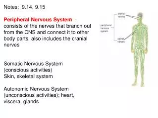
Includes all neural tissue outside the CNS The Peripheral Nervous System (PNS)
Deliver sensory information to the CNS Carry motor commands to peripheral tissues and systems Functions of the PNS
Also called peripheral nerves: • bundles of axons with connective tissues and blood vessels • carry sensory information and motor commands in PNS: • cranial nerves—connect to brain • spinal nerves—attach to spinal cord Nerves
Receptors: • detect changes or respond to stimuli • neurons and specialized cells • complex sensory organs (e.g., eyes, ears) • Effectors: • respond to efferent signals • cells and organs Receptors and Effectors
Afferent division: • carries sensory information • from PNS sensory receptors to CNS • Efferent division: • carries motor commands • from CNS to PNS muscles and glands Functional Divisions of the PNS
Somatic nervous system(SNS) Autonomic nervous system (ANS) The Efferent Divisions of the PNS
Includes all somatic motor neurons that innervate skeletal muscles • Controls skeletal muscle contractions: • voluntary muscle contractions • involuntary muscle contractions (reflexes) The Somatic Nervous System(SNS)