Download
1 / 17
330 likes | 2.69k Vues
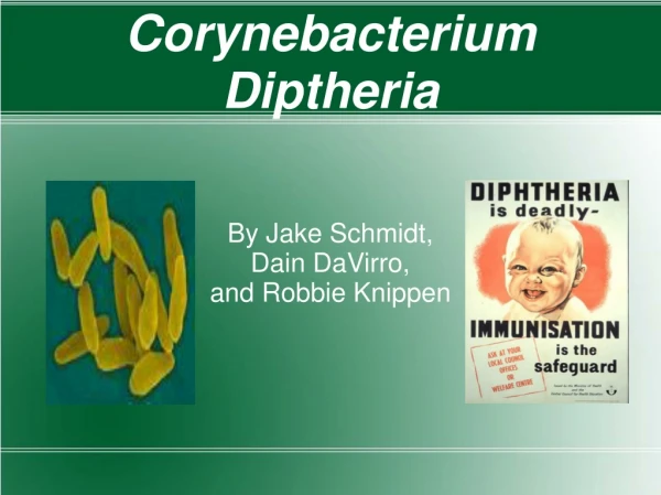
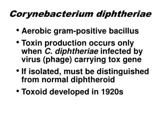
Corynebacterium diphtheriae. By. Dr. Emad AbdElhameed Morad. Lecturer of Medical Microbiology and Immunology. Morphology. Gram positive bacilli arranged in acute angled pairs (Chineese letter arrangement). The bacilli are swollen at one end (club shaped bacilli).

E N D
Corynebacterium diphtheriae By Dr. Emad AbdElhameed Morad Lecturer of Medical Microbiology and Immunology
Morphology Gram positive bacilli arranged in acute angled pairs (Chineese letter arrangement). The bacilli are swollen at one end (club shaped bacilli). The bacilli are beaded due to the metachromatic volutin granules. These volutin granules could be visualized by methylene blue stain.The granules appear dark blue while the body of the bacillus stains light blue.
Diphtheroids by Gram stain Diphtheriods are normal flora in the throat, conjunctiva and on the skin. They are similar in morphology to Corynebacterium diphtheria.
Culture Aerobe. Grow best on Loffler’s serum at 37 degree. It can grow on blood tellurite medium (blood agar + 0.04% potassium tellurite) on which it gives grey to black colonies.
Toxigenicity tests Elek’s test: A strip of filter paper saturated with diphtheria antitoxin is embedded in the agar plate. Diphtheria is inoculated at right angles to the filter paper, then plates are incubated for two days at 37 degree. If the organism is toxigenic, fine white lines of precipitation occur commencing from the streak.
In-vivo test: Injecting two guinea pigs with the diphtheria isolate. If the guinea pig protected with the antitoxin survives, while the unprotected one dies, the isolate is considered toxigenic.
PCR: For detection of the diphtheria toxin genes. ELISA: For detection of the diphtheria toxin. Tissue culture test: The test strain is inoculated into an agar overlay of cell culture. The toxin produced diffuses into the cells below and kill them.
Laboratory diagnosis Specimen:throat swab in faucial diphtheria. Direct smears stained with Gram stain and methylene blue: Gram positive bacilli + Chineese letter arrangement + beaded appearance. Culture:is done on Loffler’ serum and blood tellurite media for 12-18 hours. Identificationof the bacteria on culture is done by: Staining by Gram and MB that must be confirmed by Toxigenicity tests (the most definitive for diagnosis).