Download

1 / 52
550 likes | 1.21k Vues
BASIC ANATOMICAL TERMINOLOGY. ANATOMICAL POSITION. The anatomical position is a standardized method of observing or imaging the body that allows precise and consistent anatomical references. . ANATOMICAL POSITION.

E N D
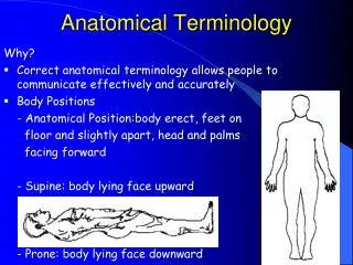
ANATOMICAL POSITION • The anatomical position is a standardized method of observing or imaging the body that allows precise and consistent anatomical references.
ANATOMICAL POSITION • When in the anatomical position, the subject stands erect facing the observer, the upper extremities are placed at the sides, the palms of the hands are turned forward, and the feet are flat on the floor.
THE ANATOMICAL POSITION
TERMINOLOGY • Reclining Position • If the body is lying face down, it is in the prone position. • If the body is lying face up, it is in the supine position.
REGIONAL NAMES • Are names given to specific regions of the body for reference. • Examples: include cranial (skull), thoracic (chest), brachial (arm), patellar (knee), cephalic (head), and gluteal (buttock)
PLANES • Planes are imaginary flat surfaces that are used to divide the body or organs into definite areas & include: • Midsagittal (medial) and parasagittal, frontal (coronal), transverse (cross-sectional or horizontal) and oblique.
SECTIONS • Sections are flat surfaces resulting from cuts through body structures. They are named according to the plane on which the cut is made and include transverse, frontal, and midsagittal
Horizontal (or cross) section Frontal (or coronal) plane Saggital plane
DIRECTIONAL TERMS • Directional terms are used to precisely locate one part of the body relative to another and to reduce length of explanations.
DIRECTIONAL TERMS • Superior/Cephalic/Cranial • Inferior/Caudal • Anterior/Ventral/Rostral • Posterior/Dorsal • Superficial: toward surface • Deep: away from surface
DIRECTIONAL TERMS • Medial: toward midline • Lateral: away from midline • Intermediate: between 2 points • Ipsilateral: same side • Contralateral: opposite side • Proximal: near origin • Distal: away from origin
DIRECTIONAL TERMS • External (Outer) • Internal (Inner) • Central • Peripheral • Parietal • Visceral
AREAS • Head & Neck • Trunk • Thorax • Abdomen • Pelvis & Perineum • Extremities (or limbs) • Upper • Lower
BODY CAVITIES • Cranial • Thoracic • Abdominal • Pelvic
BODY CAVITIES • Body Cavities - Body cavities are spaces within the body that help protect, separate, and support internal organs. • Dorsal Body Cavity • Ventral Body Cavity
BODY CAVITIES • Dorsal Body Cavity - The dorsal body cavity is located near the dorsal (back) surface of the body and has two subdivisions, the cranial cavity and the vertebral canal.
BODY CAVITIES • The cranial cavity is formed by the cranial bones and contains the brain.
BODY CAVITIES • The vertebral (spinal) canal is formed by the bones of the vertebral column and contains the spinal cord. • Three layers of protective tissue, called meninges, line the dorsal body cavity.
BODY CAVITIES • Ventral Body Cavity - The ventral body cavity is subdivided by the diaphragm into an upper thoracic cavity and a lower abdominopelvic cavity.
BODY CAVITIES • The thoracic cavity contains two pleural cavities, and the mediastinum, which includes the pericardial cavity.
UPPER THORACIC CAVITY • The pleural cavities enclose the lungs, while the pericardial cavity surrounds the heart.
UPPER THORACIC CAVITY • The mediastinum is a broad, median partition between the lungs that extends from the sternum to the vertebral column, it contains all contents of the thoracic cavity except the lungs. • The pericardial cavity encloses the heart and great vessels.
ABDOMINOPELVIC CAVITY • The abdominopelvic cavity is divided into a superior abdominal and an inferior pelvic cavity.
ABDOMINOPELVIC CAVITY • Viscera of the abdominal cavity include the stomach, spleen, pancreas, liver, gallbladder, small intestine, and most of the large intestine
ABDOMINOPELVIC CAVITY • Viscera of the pelvic cavity include the urinary bladder, portions of the large intestine and internal female and male reproductive structures.
ABDOMINOPELVIC CAVITY • Thoracic and AbdominalCavityMembranes: • A thin, slippery serous membrane covers the viscera within the thoracic and abdominal cavities and also lines the walls of the thorax and abdomen.
ABDOMINOPELVIC CAVITY • Parts of the serous membrane are the parietal layer which lines the walls of the cavities and the visceral layer which covers and adheres to the viscera within the cavities.
ABDOMINOPELVIC CAVITY • Serous fluid between the two layers reduces friction and allows the viscera to slide somewhat during movements. • The serous membranes include the pleura, pericardium and peritoneum.
PLEURALMEMBRANE • The pleuralmembrane surrounds the lungs, with the visceral pleura clinging to the surface of the lungs and the parietal pleura lining the chest wall.
PERICARDIUM • The serous membrane of the pericardial cavity is the pericardium, with visceral pericardium covering the surface of the heart and the parietal pericardium lining the chest wall.
PERITONEUM • The peritoneum is the serous membrane of the abdominal cavity, with the visceral peritoneum covering the abdominal viscera and the parietal peritoneum lining the abdominal wall.
ABDOMINOPELVIC REGIONS • To describe the location of organs easily, the abdominopelvic cavity may be divided into nine regions by drawing four imaginary lines
ABDOMINOPELVIC QUADRANTS • To locate the site of an abdominopelvic abnormality in clinical studies, the abdominopelvic cavity may be divided into quadrants by passing imaginary horizontal and vertical lines through the umbilicus.