Download
1 / 46
460 likes | 474 Vues
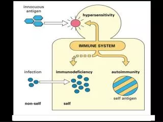
Hypersensitivity ( 超敏反应 ). Qingqing Wang Institute of Immunology Zhejiang University School Of Medicine wqq@zju.edu.cn. Type II hypersensitivity (cytotoxic type).

E N D
Hypersensitivity (超敏反应) Qingqing Wang Institute of Immunology Zhejiang University School Of Medicine wqq@zju.edu.cn
Type IIhypersensitivity(cytotoxic type) • Type IIhypersensitivity reactions are mediated by IgG and IgM antibody binding to specific cells or tissues. The damage caused is thus restricted to the specific cells or tissues bearing the antigen. • The antibodies damage cells and tissues by activating complement, and by binding and activating effector cells carrying FcR.
I. Pathogenic mechanisms Ags on the surface of target cells ↓ body→IgG, IgM ↓ 1. damage the target cell 1)activation of complement 2)opsonization: FcR, C3bR 3) ADCC: NK cells, M 2. target cell dysfunction
NK cell opsonization
Effector mechanisms of Ab-mediated diseases In myasthenia gravis the Abs against Ach receptor inhibit neuromuscular transmission and cause paralysis Graves’ disease
II. Clinical disease 1.Transfusion reactions A型血输入B型血体内 B型血体内存在抗A抗体 输血 A型血 抗原 抗体 反应 A型血抗原 活化 血细胞溶解 补体
2. Hemolytic disease of the newborn Mainly occurs when an Rh- mother gives birth to an Rh+ infant. Prevention: The administration of anti-Rh Ab to an Rh- mother within 72 hours of delivering an Rh+ infant will prevent sensitization and problems with subsequent pregnancies.
3.Autoimmune hemolytic disease Some drugs or viruses stimulate Ab formation by changing the erythrocyte surface components to form new epitopes. The resulting Abs can cross react with epitopes on unmodified RBCs.
4.Drug-induced reaction to blood components These diseases have been associated with many different chemotherapeutics, such as penicillin, the sulfonamides. These drugs stimulate Ab formation by forming new epitopes with serum proteins, which then adsorb nonspecifically to the red blood cell surface so that its new epitopes are expressed.
5. Graves’ disease The patients produce antibodies to thyrotropin (thyroid stimulating hormone, TSH) receptor. The end result is overproduction of thyroid hormone and hyperthyroidism.
Type IIIhypersensitivity(immune complex type) Immune complexes (IC) deposit in basement membranes of small blood vessels in various organs.
I. Pathogenic mechanisms Ag→body→IgG, IgM, IgA ↓ immune complexes (IC) ↓ soluble IC ↓ ICs are deposited from the circulation into vascular basement membranes ①↓ ② ↓FcR activation of complement plat. and basophils ↓ C3a, C5a →mast cell → release of vasoactive amines ↓ basophils ③ Neutrophils vasodilation ↓ lysosomal edema enzymes→damage the tissue aggravate
IC are capable of triggering a variety of inflammatory processes • ICs interact with the complement system to generate C3a and C5a. These complement fragments stimulate the release of vasoactive amines and are chemotactic factors for mast cells and basophils, eosinophils and neutrophils. • ICs interact directly with basophils and platelets (via FcR) to induce the release of vasoactive amines. • Neutrophils exocytose their lysosomal enzymes onto the site of IC deposition and damage the underlying tissue.
Deposition of IC in tissues • An increase in vascular permeability In general, complement, mast cells, basophils and platelets must all be considered as potential producers of vasoactive amines. • Local high blood pressure and turbulence The blood pressure in the glomerular capillaries is approximately four times that of most other capillaries.
II. Clinical diseases 1. Arthus reaction An animal is immunized repeatedly until it has appreciable levels of serum Ab (mainly IgG). Following subcutaneous or intradermal injection of the antigen a reaction develops at the injection site, sometimes with marked edema and hemorrhage.
Arthus’s reaction (1903) 经抗原反复免疫之后,注射抗原的皮下出现局部红肿、出血和坏死等剧烈炎症反应。
基底膜 2 内皮细胞 抗原 免疫复合物沉淀 抗体 中性粒细胞 补体 复合物 C3a C5a 血小板凝聚 血小板 C5a 肥大细胞 趋化 微血栓形成 血管活性胺 活性酶 血管壁 1
2. Serum sickness Serum sickness is a complication of serum therapy, in which massive doses of anti-serum are given in conditions such as snake bite.
Serum sickness(血清病) Clemens Pirquet 1874-1929
3. Postinfectious glomerulonephritis 常见于A族溶血性链球菌感染后2-3周。抗体与链球菌可溶性抗原形成复和物,沉积于肾小球基底膜处
4.Rheumatoid arthritis • Rheumatoid factor (RF):an immunoglobulin (mainly IgM but also IgG and IgA) with antibody specificity for the Fc portion of IgG. • The joint synovial fluid contains IC consisting of RF-IgG-complement. • Many patients with rheumatoid arthritis also have antinuclear antibodies. 5. Systemic lupus erythematosus (SLE) • antinuclear antibodies • hypergammaglobulinemia
RA,SLE 自身抗体与可溶性自身抗原形成免疫复和物,沉积于皮下、关节和肾小球基底膜等处。
Type IVhypersensitivity(Delayed type hypersensitivity) • Delayed type hypersensitivity is initiated by sensitized T cells reacting with specific antigens. The reactions are manifest as inflammation at the site of antigen exposure, which usually peaks 24-72 hours after exposure. • This reaction is independent of antibody and complement.
I. Pathogenic mechanisms Ag-MHC Antigen→APC→T cells co-stimulating factors↓ sensitized T cell ↓ effector and memory cells ↓ ↓ CD4+T cell (Th1 type) CD8+T cell (CTL) ↓ ↓ release of cytokines killing target cells ↓ by the release of inducing the inflammatory perforin and granzymes response or by the FasL-Fas pathway (primarily M and T cells)
II. Clinical diseases 1. Infectious DTH In the infective process, intracellular parasitical bacteria (Mycobacterium tuberculosis, Mycobacterium leprae, Brucella), viruses and fungi cause T cell-mediated immune responses, which are referred to as infectious delayed type hypersensitivity.
Tuberculin-type hypersensitivity The tuberculin skin test (OT) reaction principally involves M tuberculin→body→T cells are activated ↓ IFN-→M→TNF, IL-1 ↓ endothelial cells in dermal blood vessels ↓express CAM: E-selectin, ICAM-1, VCAM-1 ↓ recruiting monocytes and T cells (Monocytes constitute 80-90% of the total cellular infiltrate)
Tuberculin-like delayed type hypersensitivity reaction are used practically in two ways. 1) To confirm past infection with M. tuberculosis, but not necessarily active disease. 2) To be a general measure of cell-mediated immunity.
2. Contact dermatitis • Langerhans cells and keratinocytes acting as APCs have key roles in contact hypersensitivity. • Keratinocytes produce a range of cytokines. • A contact hypersensitivity reaction has two stages: sensitization and elicitation. Sensitization produces a population of memory T cells and elicitation involves recruitment of CD4+ lymphocytes and macrophages.
接触羽毛 接触橡胶
Many important sensitizing allergens are organic chemicals, and some are metals such as nickel, chromate. It is assumed that they function as haptens. • When allergen again penetrates the skin, these memory cells rapidly evolve into effectors that mediate a delayed-type hypersensitivity reaction at the site of penetration.
Thanks for your attention! • Thank you very much for your support and cooperation in my teaching • If you have any question and suggestion, please feel free to contact me: wqq@zju.edu.cn