Download
1 / 3
30 likes | 45 Vues
DenseBreast-info.org is an online education resource for dense breast information. You can earn a lot of knowledge for dense breast tissue, facts, screening tests etc.

E N D
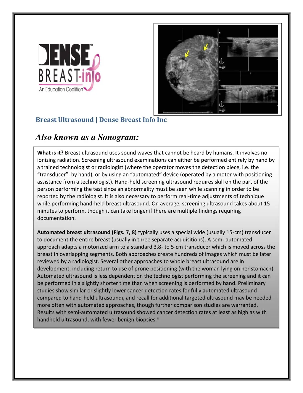
Breast Ultrasound | Dense Breast Info Inc Also known as a Sonogram: What is it? Breast ultrasound uses sound waves that cannot be heard by humans. It involves no ionizing radiation. Screening ultrasound examinations can either be performed entirely by hand by a trained technologist or radiologist (where the operator moves the detection piece, i.e. the “transducer”, by hand), or by using an “automated” device (operated by a motor with positioning assistance from a technologist). Hand-held screening ultrasound requires skill on the part of the person performing the test since an abnormality must be seen while scanning in order to be reported by the radiologist. It is also necessary to perform real-time adjustments of technique while performing hand-held breast ultrasound. On average, screening ultrasound takes about 15 minutes to perform, though it can take longer if there are multiple findings requiring documentation. Automated breast ultrasound (Figs. 7, 8) typically uses a special wide (usually 15-cm) transducer to document the entire breast (usually in three separate acquisitions). A semi-automated approach adapts a motorized arm to a standard 3.8- to 5-cm transducer which is moved across the breast in overlapping segments. Both approaches create hundreds of images which must be later reviewed by a radiologist. Several other approaches to whole breast ultrasound are in development, including return to use of prone positioning (with the woman lying on her stomach). Automated ultrasound is less dependent on the technologist performing the screening and it can be performed in a slightly shorter time than when screening is performed by hand. Preliminary studies show similar or slightly lower cancer detection rates for fully automated ultrasound compared to hand-held ultrasoundi, and recall for additional targeted ultrasound may be needed more often with automated approaches, though further comparison studies are warranted. Results with semi-automated ultrasound showed cancer detection rates at least as high as with handheld ultrasound, with fewer benign biopsies.ii
Automated Breast Ultrasound Coronal view (left), transverse view (top right), and sagittal view (bottom right) images from automated US show two irregular hypoechoic (dark gray) masses due to grade 2-3 invasive ductal carcinoma (yellow arrows). How it works: Ultrasound uses high-frequency sound waves to form an image (sonogram). The sound waves pass through the breast and bounce back or “echo” from various tissues to form a picture of the internal structures of the breast. Only gentle pressure is applied to the breasts and ultrasound rarely causes any discomfort. Ultrasound does not use or produce ionizing radiation. A water-soluble gel or lotion is placed on the skin of the breast. A hand-held device (transducer) directs the sound waves to the breast tissue. The transducer is moved over the skin of the breast to create a picture that can be seen on a computer screen. Cancers are usually seen as masses that are slightly darker than the normal lighter gray fat or white (fibrous) breast tissue (Figs 9, 10). Sometimes distortion of the tissue or bright (white) echogenic dots due to calcifications can be seen. Cysts are round or oval black fluid-filled sacs and are often seen with ultrasound; cysts are a normal finding (Fig 11). Some ultrasound equipment also allows assessment of tissue stiffness through use of elastography; this can be used to help determine need for biopsy of low suspicion lesions, with soft lesions more likely benign and stiff lesions more likely malignantiii, iv. Figure 9. Ultrasound of Cancer. This 60-year-old woman was noted to have an irregular mass on screening mammography. Ultrasound (US) shows an irregular, hypoechoic (dark gray) spiculated mass (arrow), highly suspicious for cancer. US-guided biopsy and subsequent surgery showed invasive lobular cancer.
Ultrasound of Triple Negative Breast Cancer :- This 32-year-old woman was 10 weeks pregnant and noted a lump in her left breast. US showed an oval hypoechoic (dark gray) mass (white arrow) with surrounding hyperechoic (whiter) rim (short yellow arrows). Possibilities included abscess and cancer. US-guided fine needle aspiration did not show pus, so core biopsy was performed, showing grade 3 invasive ductal cancer, lacking estrogen receptors (ER), progesterone receptors (PR), or human epidermal growth factor 2 (HER2) receptors, i.e. an aggressive subtype of breast cancer called “triple negative” breast cancer. Such cancers can sometimes be difficult to distinguish from a cyst. Ultrasound of a simple cyst :- This 73-year-old woman uses estrogen cream and a new mass was seen on her screening mammogram. On ultrasound targeted to the mammographic mass, a circumscribed (well-defined) oval anechoic (black) mass is seen, with increased echoes (whiter) deep to the mass (called “posterior enhancement”), i.e. a simple cyst, which is a benign finding. Cysts do not require follow-up or biopsy. When very large, a cyst can be aspirated (drained) if it is causing pain. Benefits: Physician-performed ultrasound finds an additional 3 to 4 cancers per thousand women already screened by mammographyv,vi; or tomosynthesis (3D mammography)vii automatedviii or technologist-performedix ultrasound finds 2 to 3 cancers per thousand women screened. More than 85% of cancers seen only on ultrasound are invasive, early stage, and node negative. In a prospective randomized study from Japanx, women who had screening ultrasound in addition to mammography were half as likely to have cancer detected because of a lump or other symptoms (interval cancer) before the next screen. Ultrasound is readily available and cost effective, though not all centers offer screening ultrasound due to a shortage of trained personnel. Considerations: Performing ultrasound requires experience and expertise of both the individual performing the scanning (technologist and/or radiologist) and of the radiologist who interprets the images. On average, ultrasound will show more areas which need follow-up than does mammography. Some of those “finds” will be cancer, but the vast majority, determined after further imaging or biopsy, will not be cancer (known as a “false positive”). In two multicenter prospective trialsv,x, 20-30% of cancers were seen only on mammography and 29-33% of cancers were seen only on ultrasound: it is important to continue mammography in addition to ultrasound screening. Contact As DenseBreast-info, Inc. PO Box 997 Deer Park, NY 11729 http://densebreast-info.org