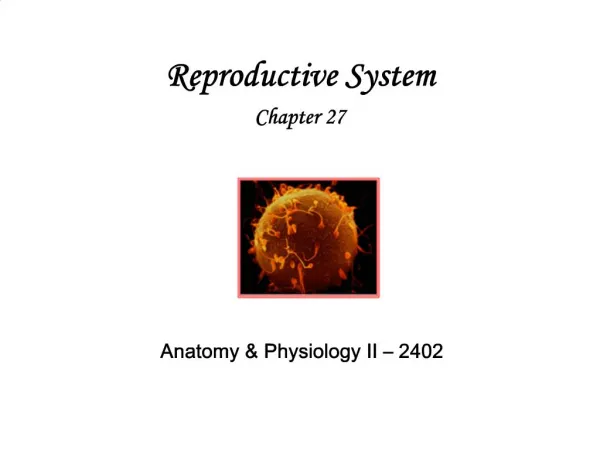
Download
1 / 23
230 likes | 422 Vues
CLINICAL ANATOMY II. COMMITTEEE. 17. December.2013 Tuesday. Kaan Yücel M.D., Ph.D. INTRORUCTION TO OSTEOLOGY. The Two Comedians , by Chris Peters. Kaan Yücel M.D., Ph.D. 19. November 201 3 Tuesday. Accessory Bones.

E N D
CLINICAL ANATOMY II. COMMITTEEE 17. December.2013 Tuesday Kaan Yücel M.D., Ph.D.
INTRORUCTION TO OSTEOLOGY TheTwoComedians, byChrisPeters Kaan Yücel M.D., Ph.D. 19. November2013 Tuesday
Accessory Bones • Accessory (supernumerary) bones develop when additional ossification centers appear and form extra bones. • Many bones develop from several centers of ossification, and the separate parts normally fuse. • Sometimes one of these centers fails to fuse with the main bone, giving the appearance of an extra bone. .
Heterotopic Bones • Bones sometimes form in soft tissues where they are not normally present (e.g., in scars). • Horse riders often develop heterotopic bones in their thighs (rider's bones), probably because of chronic muscle strain resulting in small hemorrhagic (bloody) areas that undergo calcification and eventual ossification. .
Changes in Bones & Bone Fractures • Trauma to a bone may break it. For the fracture to heal properly, the broken ends must be brought together, approximating their normal position. reduction of a fracture. • Fractures are more common in children than in adults. .
Changes in Bones & Bone Fractures • Immediately after a fracture, the patient suffers severe local pain and is not able to use the injured part. • Deformity may be visible if the bone fragments have been displaced relative to each other. .
OSTEOPOROSIS decreases in theorganic & inorganiccomponents of the bone byaging . • Bones become brittle, lose their elasticity, and fracture easily. • Bone scanning is an imaging method used to assess normal and diminished bone mass.
(BONE) SCINTIGRAPHY metabolic activity of bone and its affinity to uptake a detectable marker image can be captured by a scan a wide range of indications ranging from sports related injuries to detection of metastasis (spreading of cancer) to the bones. .
BONE DENSITOMETRY (DEXA, DXA) enhanced form of x-ray technology used to measure bone loss . • most often used to diagnose osteoporosis • effective in tracking the effects of treatment for osteoporosis and other conditions that cause bone loss.
SKULL BONES View of a Skull, 1489by Leonardo Da Vinci 5.10.2012 Kaan Yücel M.D., Ph.D.
HEAD INJURIES • major cause of death and disability • complications • Hemorrhage • Infection • Injury to the brain and cranial nerves
FRACTURES OF THE CRANIAL FOSSAE • In fractures of the anterior cranial fossa, the cribriform plate of the ethmoid bone may be damaged. • Fractures of the middle cranial fossa are common, because this is the weakest part of the base of the skull.
VERTEBRAL COLUMN, RIBS & STERNUM byIsabella Kung 10. December.2013 Tuesday Kaan Yücel M.D., Ph.D.
Scoliosis Greekskoliōsisskolios"crooked" spine is curvedfromsidetoside 2% of women,lessthan0.5 % of men. Progressivedisease Originunknown (idiopathic) 80% of thecases, evidencefor a geneticandnutritionalcomponent
Scoliosis Oftenincludes a twisting of the spine, resulting in distortion of the ribs and entire thorax. Usuallypresents in pre-teens and adolescents. Structural scoliosis may require surgical intervention; alternatively scoliosis may be corrected using orthotics (e.g. braces).
Hyperkyphosis Kyphosis natural curvatures of the thoracic spine Hyperkyphosisa pathologically exaggerated thoracic curvature, commonly called "hunchback." Commonin aging adults, usually aided by the vertebral collapse related to osteoporosis. Other common causes trauma, arthritis, and endocrine or other diseases.
Hyperlordosis Lordosisnaturalcurvature of thelumbarspine hyperlordosisis a pathologicallyexaggeratedlumbarcurvature, commonlycalled "swayback.« Symptomsmayincludepainandnumbnessifthenervetrunksarecompromised.
Hyperlordosis Attributedtoweakbackmusclesor a habitualhyperextension, such as in pregnantwomen, men withexcessivevisceralfat, andsomedancepostures. Alsocorrelatedwithpuberty.
RIB FRACTURES • The short, broad 1st rib, rarely fractured • When broken ---structures crossing its superior aspect injured, including the brachial plexus of nerves and subclavian vessels. • The middle ribs most commonly fractured. • The weakest part of a rib is just anterior to its angle.
SupernumeraryRibs Thenumber of ribsincreasedbythe presence of cervicaland/orlumbarribs Cervicalribsrelativelycommon (0.5-2%) interferewithneurovascularstructuresexitingthesuperiorthoracicaperture. Supernumerary (extra) ribs Clinicalsignificanceconfusion in radiologicaldiagnosis Supernumerary ribs in a neonate 14 pairs of ribs in thechest X-ray
SternalFractures • Despite the subcutaneous location of the sternum, sternal fractures are not common.Airbag • A fracture of the sternal body is usually a comminuted fracture (a break resulting in several pieces). • The most common site in elderly people @ the sternal angle • The concern in sternal injuries heart injury or lung injury
MedianSternotomy • Togainaccesstothethoraciccavityforsurgicaloperations in themediastinum—e.g., coronaryartery bypass grafting—thesternum is divided (split) in themedianplaneandretracted. • A goodexposureforremoval of tumors in thesuperiorlobes of thelungs. • Aftersurgery, thehalves of thesternumarejoinedusingwiresutures.
SternalAnomalies Complete sternalcleftuncommonanomalythroughwhichtheheartmayprotrudeectopiacordis PartialcleftsSternalforamen A receding (pectusexcavatum, orfunnelchest)orprojecting (pectuscarinatum, orpigeonbreast) sternum