Download

1 / 58
710 likes | 1.39k Vues
Marc L. Braithwaite, OD Vision Care of Maine. Keratoconus And specialty contact lens fitting of irregular corneas. Keratoconus. What have the years taught us?. Keratoconus Characteristics. Non-inflammatory. Central or para -central corneal thinning. Corneal steepening or protrusion.

E N D
Marc L. Braithwaite, OD Vision Care of Maine KeratoconusAndspecialty contact lens fitting of irregular corneas
Keratoconus • What have the years taught us?
Keratoconus Characteristics • Non-inflammatory. • Central or para-central corneal thinning. • Corneal steepening or protrusion. • Increased astigmatism and possibly myopia. • Loss of best spectacle corrected visual acuity. • Corneal striae and scarring. • Corneal hydrops (inflammatory).
Pathology of Keratoconus • Loss of Bowman’s Layer. • Stromal Thinning. • Apoptosis. • Increased Enzyme Activity. • Enlarged Prominent Corneal Nerves.
Causes of Keratoconus • Heredity vs. Mechanical • Cellular • Tissue • Genetic
Heredity vs. Mechanical • Does eye rubbing cause Keratoconus? • 2 out of 250 doctors feel that rubbing is a cause. • KC patients do rub their eyes more often than those without KC. • What is it that makes KC patients rub their eyes?
Cellular Changes • Keratoconus cells are hypersensative. • Increased enzyme activity, lack of enzyme inhibitors. • Matrix substrate instability in response to environmental stress factors. • mtDNA damage and exaggerated oxidative response causing cellular damage.
Tissue Changes • Loss of Bowman’s layer. • Lamellar slippage. • Lack “anchoring” lamellar fibrils. • Apoptosis of the stroma causing anterior thinning.
Genetics • Autosomal dominant w/variable penetrance. • SOD1, an antioxidant enzyme, is abnormal in some KC corneas. • No single gene responsible. • 10 different chromosomes have been associated with KC. • Most likely multiple genes involved.
Additional Information • Male to Female Ratio = 3:1 • Approximately 20% result in PKP. • 90% are diagnosed by optometrists. • Mean age of diagnosis is 22.88 years. • Visual outcome with RGP is better than PKP. • More prevalent in certain ethnic groups (4x higher in Asians from Indian sub-continent regions than White Europeans).
Progression and Prognosis • Age is a big factor. • The younger the diagnosis, the poorer the prognosis. • Less likely to progress to the point of a transplant if diagnosed in the 30’s. • 20% of Keratoconus patients result in corneal transplants. • 35 to 45% of all transplants are due to Keratoconus.
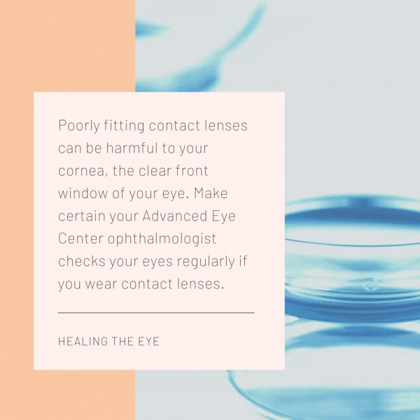
Possible Aggravating Factors • UV exposure. • Allergies. • Vigorous eye rubbing. • Poorly fitting contact lenses. • Inflammation.
Types of Keratoconus • Nipple/Oval cone - central or mildly para-central localized thinning and steepening. • Keratoglobus - Large generalized thinning and steepening. • PMD (pellucid marginal degeneration) – peripheral thinning and steepening. • Keratoconus Fruste – Less progressive and less manipulative.
Nipple/Oval Cone • Central Steepening • Steepest form
Keratoglobus • Wider – 75 to 90% of cornea. • Not as steep.
Pellucid Marginal Degeneration • Peripheral Thinning
How to Treat Keratoconus • Spectacles • Contacts • Soft Standard • Soft Custom • RGP Standard • RGP Custom • Hybrid • Surgery • Intacs • Penetrating Keratoplasty • Riboflavin/UV treatment
When to Intervene? • Best Spectacle/Soft CL Acuity 20/30 or better? • Good tolerance of acuity. • Corneal health is not compromised. • “If it aint broke, don’t fix it.” • Best Spectacle/Soft CL Acuity worse than 20/30? • Specialized contact lenses. • My opinion, use RGP lenses.
Which RGP Design? • Early Keratoconus • Standard RGP • KC RGP • Mid-stage Keratoconus • KC RGP • Custom KC RGP • Advanced Keratoconus • Custom KC RGP • Intra-limbal or Scleral RGP
My “GO TO” Lens – Rose K • Developed by Dr. Paul Rose. • Designed to fit the irregular cornea. • “Very forgiving lens” • Multiple designs to fit all shapes of corneas and corneal conditions. • Blanchard is very good to work with and has staff to assist with very difficult cases.
Nipple/Oval Cone Fitting • Most common form of KC. • Early stages - simple RGP or KC RGP • Later stages – KC RGP usually small and steep. • The steeper the cone, the smaller the lens diameter.
Rose K2 • Rose K vs. Rose K2 • 72% of patients notice an increase in acuity with aspheric, aberration control. • Lens to be centered on the cone. • Reduce excessive movement (1 to 2mm).
Fitting the Rose K2 • Too high – tighten edge lift reduce OAD steepen base curve • Too low – increase edge lift increase OAD flatten base curve
Fitting the Rose K2 • Centrally fitting the lens on a nipple cone better insures optimal acuity and comfort.
Rose K2IC • IC stands for irregular cornea • Larger diameter • Larger optic zone • Aspheric for aberration control • Reverse geometry design
PMD • Keratoglobus • LASIK induced ectasia • Corneal transplants
Corneal Dystrophies • Traumatic Corneas with Scars • Post RK • Irregular Astigmatism or Corneal Warpage
Fitting with ACT Using ACT ( Asymmetric Corneal Technology) • 3 standard grades available • Option also to specify degree of tuck in 0.1 steps from 0.4 to 1.5mm Grade 3 (1.3mm steeper) Grade 1 ( 0.7mm steeper) Grade 2 (1.0mm steeper)
Fitting with ACT ACT - Improved comfort , lens stability and vision NO ACT WITH ACT
Fitting Pearls • Tendency to tighten after initial fitting. • Light central touch will increase acuity. • Avoid central staining. • Movement is necessary but slight movement is usually sufficient. • Pay attention to tear flow beneath lens. • The steeper the lens, the smaller OAD and less movement. • Don’t change too many parameters at once.
Penetrating KeratoplastyWhen to refer? • Acuity is 20/50 or worse. • Patient intolerance to visual decrease. • Scars within the visual axis. • Multiple episodes of Hydrops. • Contact lens intolerance. • Unable to get adequate/healthy CL fit. • Consider OD to OD referral. • Give reasonable expectations.
Post PKP Management • How soon can you fit with lens? • Why are the curvatures so strange? • Do you have to wait for all sutures to be removed? • Corrective options. • Spectacles • RGP contact lenses. • LASIK
Rose K2 Post Graft • Much more difficult to fit than KC. • Patients are less tolerable to CL. • Eyes are more dry. • Ill-fitting contact lenses can lead to graft rejection. • Lens design is crucial to success.
K2PG Fitting Pearls • Don’t be intimidated! • Watch tear flow! • Also good lens for ectasia patients. • Stay with your fitting basics • Fit base curves. • Adjust diameter. • Adjust peripheral curves. • Use ACT or Toric PC if needed.
The Difficult Ones • Nothing is comfortable. • Acuity isn’t improving.. • Eyes are too dry. (Sjogren’s Syndrome) • Cornea is too irregular for any lens to fit properly or in a healthy manner.