Download

1 / 13
140 likes | 246 Vues
Infectious Arthritis (S. Aureus ). INTRODUCTION.

E N D
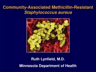
INTRODUCTION Staphylococcus aureus, the most virulent of the many staphylococcal species, has demonstrated its versatility by remaining a major cause of morbidity and mortality despite the availability of numerous effective antistaphylococcal antibiotics. S. aureus is a pluripotent pathogen, causing disease through both toxin-mediated and non-toxin-mediated mechanisms. This organism is responsible for both nosocomial and community-based infections that range from relatively minor skin and soft tissue infections primarily to life-threatening systemic infections. (Harrison’s Principles of Internal Medicine, 17 Edition Volume 1, p.872) (Harrison’s Principles of Internal Medicine , 18 Edition) Staphylococci, gram-positive cocci in the family Micrococcaceae, form grapelike clusters on Gram's stain (Picture 1 and Picture 2). These organisms are catalase-positive (unlike streptococcal species), nonmotile, aerobic, and facultatively anaerobic. They are capable of prolonged survival on environmental surfaces in varying conditions. (Harrison’s Principles of Internal Medicine, 17 Edition Volume 1, p.872-873) (Harrison’s Principles of Internal Medicine , 18 Edition) S. aureus is distinguished from other staphylococcal species by its production of coagulase, a surface enzyme that converts fibrinogen to fibrin. Latex kits designed to detect both protein A and clumping factor also distinguish S. aureus from other staphylococcal species. S. aureus ferments mannitol, is positive for protein A, and produces DNAse. On blood agar plates, S. aureus tends to form golden -hemolytic colonies. (Harrison’s Principles of Internal Medicine, 17 Edition Volume 1, p.873) (Harrison’s Principles of Internal Medicine , 18 Edition)
S. aureus is distinguished from other staphylococcal species by its production of coagulase, a surface enzyme that converts fibrinogen to fibrin. Latex kits designed to detect both protein A and clumping factor also distinguish S. aureus from other staphylococcal species. S. aureus ferments mannitol, is positive for protein A, and produces DNAse. On blood agar plates, S. aureus tends to form golden -hemolytic colonies. A simple strategy for identification of the more clinically important species is outlined in Picture 3. (Harrison’s Principles of Internal Medicine, 17 Edition Volume 1, p.873) (Harrison’s Principles of Internal Medicine , 18 Edition) S. aureus is a part of the normal human flora; ~25–50% of healthy persons may be persistently or transiently colonized. The rate of colonization is higher among insulin-dependent diabetics, HIV-infected patients, patients undergoing hemodialysis, and individuals with skin damage. The anterior nares are a frequent site of human colonization, although the skin (especially when damaged), vagina, axilla, perineum, and oropharynx may also be colonized. These colonization sites serve as a reservoir of strains for future infections, and persons colonized with S. aureus are at greater risk of subsequent infection than are noncolonized individuals. (Harrison’s Principles of Internal Medicine, 17 Edition Volume 1, p.873) (Harrison’s Principles of Internal Medicine , 18 Edition)
Overall, S. aureus is a leading cause of nosocomial infections. It is the most common cause of surgical wound infections and is second only to CoNS as a cause of primary bacteremia. Increasingly, nosocomial isolates are resistant to multiple antibiotics. In the community, S. aureus remains an important cause of skin and soft tissue infections, respiratory infections, and (among injection drug users) infective endocarditis. The increasing prevalence of home infusion therapy is another cause of community-acquired staphylococcal infections. (Harrison’s Principles of Internal Medicine, 17 Edition Volume 1, p.873) (Harrison’s Principles of Internal Medicine , 18 Edition) In both children and adults, S. aureus is the most common cause of septic arthritis in native joints. This infection is rapidly progressive and may be associated with extensive joint destruction if left untreated. In adults, arthritis may result from trauma, surgery, or hematogenous dissemination. The most commonly involved joints include the knees, shoulders, hips, and phalanges. (Harrison’s Principles of Internal Medicine, 17 Edition Volume 1, p.876) (Harrison’s Principles of Internal Medicine , 18 Edition) Staphylococcus aureusis one of the most common causes of acute infectious monoarthritis. Since acute bacterial infection can destroy articular cartilage rapidly, all inflamed joints must be evaluated without delay to exclude noninfectious processes and determine appropriate antimicrobial therapy and drainage procedures. (Harrison’s Principles of Internal Medicine, 17 Edition Volume 2, p.2169-2170) (Harrison’s Principles of Internal Medicine , 18 Edition)
RISK FACTOR Infectious Arthritis of S. aureusfrequently develops in joints previously damaged by osteoarthritis or rheumatoid arthritis. Iatrogenic infections resulting from aspiration or injection of agents into the joint also occur. In these settings, the patient experiences increased pain and swelling in the involved joint in association with fever. (Harrison’s Principles of Internal Medicine, 17 Edition Volume 1, p.876) (Harrison’s Principles of Internal Medicine , 18 Edition) Some diseases increase the risk of S. aureus infection; diabetes, for example, combines an increased rate of S. aureus colonization and the use of injectable insulin with the possibility of impaired leukocyte function. Individuals with congenital or acquired qualitative or quantitative defects of polymorphonuclear leukocytes (PMNs) are at increased risk of S. aureus infections; this group includes neutropenic patients (e.g., those receiving chemotherapeutic agents), those with chronic granulomatous disease, and those with Job's or Chédiak-Higashi syndrome. Other groups at risk include individuals with skin abnormalities and those with prosthetic devices. (Harrison’s Principles of Internal Medicine, 17 Edition Volume 1, p.873) (Harrison’s Principles of Internal Medicine , 18 Edition) Infections after surgical procedures or penetrating injuries are due most often to S. aureus. (Harrison’s Principles of Internal Medicine, 17 Edition Volume 2, p.2170) (Harrison’s Principles of Internal Medicine , 18 Edition)
Patients with rheumatoid arthritis have the highest incidence of infective arthritis (S. aureus) because of chronically inflamed joints; glucocorticoid therapy; and frequent breakdown of rheumatoid nodules, vasculitic ulcers, and skin overlying deformed joints. Diabetes mellitus, glucocorticoid therapy, hemodialysis, and malignancy all carry an increased risk of infection with S. aureus. (Harrison’s Principles of Internal Medicine, 17 Edition Volume 2, p.2170) (Harrison’s Principles of Internal Medicine , 18 Edition)
PATHOGENESIS Bacteria enter the joint from the bloodstream; from a contiguous site of infection in bone or soft tissue; or by direct inoculation during surgery, injection, animal or human bite, or trauma. In hematogenous infection, bacteria escape from synovial capillaries, which have no limiting basement membrane, and within hours provoke neutrophilic infiltration of the synovium. Neutrophils and bacteria enter the joint space; later, bacteria adhere to articular cartilage. Degradation of cartilage begins within 48 h as a result of increased intraarticular pressure, release of proteases and cytokines from chondrocytes and synovial macrophages, and invasion of the cartilage by bacteria and inflammatory cells. Histologic studies reveal bacteria lining the synovium and cartilage as well as abscessesextending into the synovium, cartilage, and'in severe cases'subchondral bone. Synovial proliferation results in the formation of a pannus over the cartilage, and thrombosis of inflamed synovial vessels develops. Bacterial factors that appear important in the pathogenesis of infective arthritis include various surface-associated adhesins in S. aureus that permit adherence to cartilage and endotoxins that promote chondrocyte-mediated breakdown of cartilage. (Harrison’s Principles of Internal Medicine, 17 Edition Volume 2, p.2170) (Harrison’s Principles of Internal Medicine , 18 Edition)
CLINICAL MANIFESTATIONS Some 90% of patients present with involvement of a single joint'most commonly the knee; less frequently the hip; and still less often the shoulder, wrist, or elbow. Small joints of the hands and feet are more likely to be affected after direct inoculation or a bite. Among IV drug users, infections of the spine, sacroiliac joints, and sternoclavicular joints (Picture 4)are more common than infections of the appendicular skeleton. Polyarticular infection is most common among patients with rheumatoid arthritis and may resemble a flare of the underlying disease. (Harrison’s Principles of Internal Medicine, 17 Edition Volume 2, p.2171) (Harrison’s Principles of Internal Medicine , 18 Edition) The usual presentation consists of moderate to severe pain that is uniform around the joint, effusion, muscle spasm, and decreased range of motion. Fever in the range of 38.3°–38.9°C (101°–102°F) and sometimes higher is common but may not be present, especially in persons with rheumatoid arthritis, renal or hepatic insufficiency, or conditions requiring immunosuppressive therapy. The inflamed, swollen joint is usually evident on examination except in the case of a deeply situated joint such as the hip, shoulder, or sacroiliac joint. (Harrison’s Principles of Internal Medicine, 17 Edition Volume 2, p.2171) (Harrison’s Principles of Internal Medicine , 18 Edition) It presents as intense pain on motion of the affected joint, swelling, and fever. The most commonly involved joints include the knees, shoulders, hips, and phalanges. (Harrison’s Principles of Internal Medicine, 17 Edition Volume 1, p.876) (Harrison’s Principles of Internal Medicine , 18 Edition)
DIAGNOSIS Aspiration of synovial fluid an essential element in the evaluation of potentially infected joints can be performed without difficulty in most cases by the insertion of a large-bore needle into the site of maximal fluctuance or tenderness or by the route of easiest access. Ultrasonography or fluoroscopy may be used to guide aspiration of difficult-to-localize effusions of the hip and, occasionally, the shoulder and other joints. Normal synovial fluid contains <180 cells (predominantly mononuclear cells) per microliter. Synovial cell counts averaging 100,000/µL (range, 25,000–250,000/µL), with >90% neutrophils, are characteristic of acute bacterial infections. Crystal-induced, rheumatoid, and other noninfectious inflammatory arthritides usually are associated with <30,000–50,000 cells/µL; cell counts of 10,000–30,000/µL, with 50–70% neutrophils and the remainder lymphocytes, are common in mycobacterial and fungal infections. Definitive diagnosis of an infectious process relies on identification of the pathogen in stained smears of synovial fluid, isolation of the pathogen from cultures of synovial fluid and blood, or detection of microbial nucleic acids and proteins by nucleic acid amplification (NAA)–based assays and immunologic techniques. (Harrison’s Principles of Internal Medicine, 17 Edition Volume 2, p.2170) (Harrison’s Principles of Internal Medicine , 18 Edition) Aspiration of the joint reveals turbid fluid, with >50,000 PMNs/µL and gram-positive cocci in clusters on Gram's stain (Picture 1 and Picture 2). (Harrison’s Principles of Internal Medicine, 17 Edition Volume 1, p.876) (Harrison’s Principles of Internal Medicine , 18 Edition)
A focus of extraarticular infection such as a boil or pneumonia should be sought. Peripheral-blood leukocytosis with a left shift and elevation of the erythrocyte sedimentation rate or C-reactive protein level are common. (Harrison’s Principles of Internal Medicine, 17 Edition Volume 2, p.2171) (Harrison’s Principles of Internal Medicine , 18 Edition) Specimens of peripheral blood and synovial fluid should be obtained before antibiotics are administered. Blood cultures are positive in up to 50–70% of S. aureusinfections. The synovial fluid is turbid, serosanguineous, or frankly purulent. Gram-stained smears confirm the presence of large numbers of neutrophils. Levels of total protein and lactate dehydrogenase in synovial fluid are elevated, and the glucose level is depressed; however, these findings are not specific for infection, and measurement of these levels is not necessary for diagnosis. The synovial fluid should be examined for crystals, because gout and pseudogout can resemble septic arthritis clinically, and infection and crystal-induced disease occasionally occur together. S. aureusare seen on synovial fluid smears in nearly three-quarters of nongonococcal arthritis. Cultures of synovial fluid are positive in >90% of cases. (Harrison’s Principles of Internal Medicine, 17 Edition Volume 2, p.2171) (Harrison’s Principles of Internal Medicine , 18 Edition)
Plain radiographs show evidence of soft tissue swelling, joint-space widening, and displacement of tissue planes by the distended capsule. Narrowing of the joint space and bony erosions indicate advanced infection and a poor prognosis. Ultrasound is useful for detecting effusions in the hip, and CT or MRI can demonstrate infections of the sacroiliac joint, the sternoclavicular joint, and the spine very well. (Harrison’s Principles of Internal Medicine, 17 Edition Volume 2, p.2171) (Harrison’s Principles of Internal Medicine , 18 Edition)
DIFFERENTIAL DIAGNOSIS Cellulitis, bursitis, and acute osteomyelitis, which may produce a similar clinical picture (swollen joint), should be distinguished from septic arthritis by their greater range of motion and less than circumferential swelling. (Harrison’s Principles of Internal Medicine, 17 Edition Volume 2, p.2171) (Harrison’s Principles of Internal Medicine , 18 Edition) In patient with infectious arthritis, we should find what is the organism that’s responsible for the disease.
TREATMENT In infectious arthritis case, prompt administration of systemic antibiotics and drainage of the involved joint can prevent destruction of cartilage, postinfectious degenerative arthritis, joint instability, or deformity. In suspect patient of infectious arthritis, once samples of blood and synovial fluid have been obtained for culture, empirical antibiotics should be given that are directed against the bacteria visualized on smears or the pathogens that are likely in light of the patient's age and risk factors. Initial therapy should consist of IV administration of bactericidal agents; direct instillation of antibiotics into the joint is not necessary to achieve adequate levels in synovial fluid and tissue. An IV third-generation cephalosporin such as cefotaxime (1 g every 8 h) or ceftriaxone (1–2 g every 24 h) provides adequate empirical coverage for most community-acquired infections in adults when smears show no organisms. IV vancomycin (1 g every 12 h) is used if there are gram-positive cocci on the smear. Definitive therapy is based on the identity and antibiotic susceptibility of the bacteria isolated in culture. Infections due to staphylococci are treated with oxacillin, nafcillin, or vancomycin for 4 weeks. (Harrison’s Principles of Internal Medicine, 17 Edition Volume 2, p.2171) (Harrison’s Principles of Internal Medicine , 18 Edition)