Download

1 / 44
600 likes | 1.67k Vues
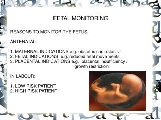
Basic Fetal Monitoring Review. Ana H. Corona, FNP-C Nursing Instructor February 2009. Electronic Fetal Monitoring. Definition of fetal monitoring Method of assessing fetal status before and during labor Why is fetal monitoring important To provide insight that may affect fetal outcomes

E N D
Basic Fetal MonitoringReview Ana H. Corona, FNP-C Nursing Instructor February 2009
Electronic Fetal Monitoring Definition of fetal monitoring Method of assessing fetal status before and during labor Why is fetal monitoring important To provide insight that may affect fetal outcomes Information is recorded on graph paper Information is permanent part of the maternal medical record Information is retrievable for litigation 11/27/2014 2
Normal Assessment Findings FHR between 110-160 in gestations 32-40+ weeks Rates slightly above 160 are normal in gestations less than 32 weeks. Regular rhythm Increases in the FHR associated with fetal movement that return to original rate range 11/27/2014 3
Electronic Fetal Monitoring Clarification Information for students is for educational purposes only Students should not assume any responsibility for interpretation of fetal monitor tracings It takes months to years of experience to be prepared to interpret fetal monitor tracings 11/27/2014 4
Methods of Electronic Fetal Monitoring • External • Noninvasive method • Utilizes an ultrasonic transducer to monitor the fetal heart • Utilizes the tocodynamometer (toco) to monitor uterine contraction pattern 11/27/2014 5
Methods of Electronic Fetal Monitoring • Internal Fetal Monitoring • Invasive • FHR is monitored via a fetal scalp electrode • Uterine activity is monitored by an intrauterine pressure catheter (IUPC) • A combination of external and internal fetal monitoring is common practice 11/27/2014 6
Advantages and Disadvantages of Internal Fetal Monitoring Advantages Patient can move without much interference in data transmission More accurate measurement of data Data less likely to be affected by artifact Disadvantages Invasive Membranes have to be ruptured and cervix dilated Application requires more skill Procedure is uncomfortable for the mother Risk of trauma and infection for mother and fetus 11/27/2014 7
Components of the Fetal Monitor Paper Tracing Strip has two components Upper graph - records FHR data Small squares represent 10 bpm increases as well as 10 seconds duration Lower graph records contraction data Small squares represent 10 second duration or 10 mmHg intensity Dark line to dark line represents one minute of time 11/27/2014 8
Baseline FHR Normal baseline FHR in a term fetus 37 completed weeks or more is 110-160 bpm. Determination of the baseline FHR is done between contractions Baseline is rounded in increments of 5 bpm example; if the FHR is running 125-135 then the baseline FHR should be documented as 130 11/27/2014 9
FHR Variability Normal changes and fluctuations in the FHR over time. Best assessed between contractions Considered to be the best indicator of fetal well-being Variability can be influenced by hypoxic events, maternal hemodynamic issues, drugs, etc. 11/27/2014 11
Examples of Variability Absent: Not detectable from baseline Minimal: Less than 5 bpm from baseline May occur with: normal fetal sleep patterns mother has received analgesia for pain Moderate : 6-25 bpm from baseline (optimal pattern) Marked: More than 25 bpm from baseline 11/27/2014 12
How Do Uterine Contractions Affect Fetal Heart Rate? • Can affect FHR by increasing or decreasing the rate in association with any given contraction. • 3 primary mechanisms by which UCs can cause a decrease in FHR • Fetal head • Umbilical cord • Uterine myometrial vessels
Periodic and Episodic FHR Characteristics Periodic: Refers to changes in the FHR that occur with or in relationship to contractions Episodic: Refers to changes in the FHR that occur independent of contractions 11/27/2014 14
Examples of Periodic Changes • Variable decelerations: Result from some type of cord compression. • Nuchal cord, True knot • Decreased amniotic fluid 11/27/2014 15
Severe Variable Decelerations Note the depth from the baseline Baseline 11/27/2014 16
Early Deceleration • Occur as a result of vagal stimulation to the fetal head during contractions which push the fetal head toward the pelvis. 11/27/2014 17
Late Decelerations • Occur in response to utero-placental insufficiency. Blood flow to the fetus is compromised and there is less oxygen available to the fetus) 11/27/2014 18
Late Decelerations with Absent Variability • Note the smoothness of the FHR pattern • Decreased FHR caused by utero-placental insufficiency • Compromised blood flow to fetus 11/27/2014 19
Prolonged Deceleration Deceleration of the FHR from the baseline lasting more than 2 minutes but less than 10 minutes. No explanation for why these occur Commonly associated with uterine hyperstimulation. Can also occur without any uterine activity 11/27/2014 20
Example Prolonged Deceleration • Note the duration of the deceleration lasts more than 2 minutes. 11/27/2014 21
FHR Accelerations Are the most common type of FHR changes Are abrupt changes and will increase from the baseline 15 bpm lasting 15 seconds before return to the baseline in a healthy gestation more than 32 weeks. Less than 32 weeks increases of 10 bpm lasting 10 seconds are indication of a well oxygenated fetus. 11/27/2014 22
Example Accelerations • Note the increase from the fetal heart baseline 11/27/2014 23
Sinusoidal Pattern • Persistent wave variation of the baseline only seen in about 2% of patients. • Related to severe fetal anemia, hypoxia, or acidosis. 11/27/2014 24
Uterine Activity Assessment Periodic tightening and relaxing of the uterine muscle. Pituitary gland is triggered to release a hormone called oxytocin that stimulates the uterine tightening. Difference in Braxton Hicks contractions and true labor is the strength of the contractions and the changes in the cervix. 11/27/2014 25
Characteristics of Contractions Frequency: How often they occur? They are timed from the beginning of a contraction to the beginning of the next contraction. Regularity: Is the pattern rhythmic? Duration: From beginning to end - How long does each contraction last? Intensity: By palpation mild, moderate, or strong. By IUPC intensity in mmHg Subjectively: Patient description 11/27/2014 26
Segments of Contractions • Increment: Beginning, building of pressure • Acme: Most intense part of the contraction • Decrement: Diminishing of the contraction • Rest: Period of time between contractions
Assessment of Contractions Palpation: Use the fingertips to palpate the fundus of the uterus Mild: Uterus can be indented with gentle pressure at peak of contraction Moderate: Uterus can be indented with firm pressure at peak of contraction Strong: Uterus feels firm and cannot be indented during peak of contraction 11/27/2014 29
Variable decelerations in FHR during labor are severe dips occurring at the peak of contraction. This FHR problem is associated with which one of the following conditions? • Utero-placental insufficiency • Fetal head compression • Uterine insufficiency • Pressure on the umbilical cord
Answer is D • These decelerations are common during labor. • The FHR drops during the contraction resulting from stimulation from chemoreceptors and baroreceptors as the cord is compressed. • The nurse should recognize these readings on the fetal monitor as normal.
A nurse is caring for a client in labor and is monitoring the FHR patterns. The nurse notes the presence of episodic accelerations on the electronic fetal monitor tracing. Which of the following actions is most appropriate? • Document the findings and tell the mother that the monitor indicates fetal well-being • Take the mothers vital signs and tell the mother that bed rest is required to conserve oxygen. • Notify the physician of the findings. • Reposition the mother and check the monitor for changes in the fetal tracing
Answer is 1 • Accelerations are transient increases in the fetal heart rate that often accompany contractions or are caused by fetal movement. • Episodic accelerations are thought to be a sign of fetal-well being and adequate oxygen reserve.
A nurse is admitting a pregnant client to the labor room and attaches an external electronic fetal monitor to the client’s abdomen. After attachment of the monitor, the initial nursing assessment is which of the following? • Identifying the types of accelerations • Assessing the baseline fetal heart rate • Determining the frequency of the contractions • Determining the intensity of the contractions
Answer is 2 • Assessing the baseline fetal heart rate is important so that abnormal variations of the baseline rate will be identified if they occur. • Options 1 and 3 are important to assess, but not as the first priority.
A nurse is monitoring a client in labor. The nurse suspects umbilical cord compression if which of the following is noted on the external monitor tracing during a contraction? • Early decelerations • Variable decelerations • Late decelerations • Short-term variability
Answer is 2 • Variable decelerations occur if the umbilical cord becomes compressed, thus reducing blood flow between the placenta and the fetus. • Early decelerations result from pressure on the fetal head during a contraction. • Late decelerations are an suggests utero-placental insufficiency during a contraction. • Short-term variability refers to the beat-to-beat range in the fetal heart rate.
The physician asks the nurse the frequency of a laboring client’s contractions. The nurse assesses the client’s contractions by timing from the beginning of one contraction: • Until the time it is completely over • To the end of a second contraction • To the beginning of the next contraction • Until the time that the uterus becomes very firm
Answer is 3 • This is the way to determine the frequency of the contractions
When monitoring the FHR of a client in labor, the nurse identifies an elevation of 15 beats above the baseline rate of 135 beats per minute lasting for 15 seconds. This should be documented as: • An acceleration • An early elevation • A sonographic motion • A tachycardic heart rate
Answer is 1 • An acceleration is an abrupt elevation above the baseline of 15 beats per minute for 15 seconds; if the acceleration persists for more than 10 minutes it is considered a change in baseline rate. • A tachycardic FHR is above 160 beats per minute.
Which of the following findings meets the criteria of a reassuring FHR pattern? • FHR does not change as a result of fetal activity • Average baseline rate ranges between 100 - 140 BPM • Mild late deceleration patterns occur with some contractions • Variability averages between 6 - 10 BPM
Answer is 4 • Variability indicates a well oxygenated fetus with a functioning autonomic nervous system. • FHR should accelerate with fetal movement. • Baseline range for the FHR is 120 to 160 beats per minute. • Late deceleration patterns are never reassuring, though early and mild variable decelerations are expected, reassuring findings.
References • AWHONN Clinical Position Statement • P. Burroughs, MSN, RN • Martin, E.J., (2002) Intrapartum Management Modules: A Perinatal Education Program. (pp 119-123). Lippincott Williams & Wilkins 3rd Edition. • Simpson, I., & Creehan, P. (2001) Perinatal Nursing 2nd Edition, (pp 379-383). Philadelphia, New York, Baltimore, Lippincott. 11/27/2014 44