Download
1 / 23
230 likes | 420 Vues
The heart and heart disease. 5.2 The cardiac cycle. Learning outcomes. Students should understand the following: Myogenic stimulation of the heart and transmission of a subsequent wave of electrical activity.

E N D
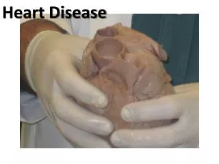
The heart and heart disease 5.2 The cardiac cycle
Learning outcomes Students should understand the following: • Myogenic stimulation of the heart and transmission of a subsequent wave of electrical activity. • Roles of the sinoatrial node (SAN), atrioventricular node (AVN) and bundle of His. • Cardiac output as the product of heart rate and stroke volume. • Pressure and volume changes and associated valve movements during the cardiac cycle. Candidates should be able to analyse and interpret data relating to pressure and volume changes during the cardiac cycle.
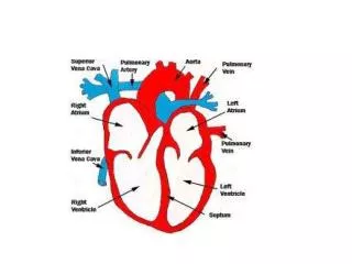
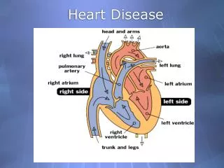
The heart • Heart muscle does not need to be stimulated by a nerve before it will contract. The heart beat originates in the muscle itself and, for this reason, it is described as being myogenic. • There are 2 phases to the beating of the heart: • Contraction (systole) • Relaxation (diastole)
The Cardiac Cycle • Cardiac cycle = sequence of events that makes up one heart beat • Continuous but can be divided into 3 stages: • Atrial systole • Ventricular systole • Ventricular diastole
1. Atrial Systole • Heart is full of blood • Contraction of both atria • AV valves open • blood flows ventricles • No backflow as veins have valves
2. Ventricular Systole • Atria relax • Ventricles contract (0.1s after atria) • Increase in pressure in ventricle • AV valves close • Semi-lunar valves open • blood flows from ventricles arteries • Lasts ~0.3 s
3. Ventricular Diastole • Ventricles relax • Blood flows from veins (at low pressure) atria • Some blood flows through atria ventricles
Atrial Systole The walls of the atria contract. This reduces the volume of the atria, increasing the pressure. More blood is forced through the atrio-ventricular valves into the ventricles. Ventricular Systole The walls of the ventricles now contract, reducing the volume in the ventricles. The pressure increases and blood is forced into the arteries. Ventricular Diastole The ventricle walls relax and the pressure in the ventricles falls. Blood starts to flow from the atria into the ventricles again.
Cardiac cycle • Cardiac cycle - narrated.swf
Control of Heartbeat • Cardiac muscle is myogenic – naturally contracts & doesn’t need to receive impulses • Need to coordinate contractions with pacemaker = sino-atrial node (SAN)
Control of heartbeat • Cardiac cycle - SAN & AVN.swf
The Sino-atrial Node • Patch of specialised muscle in wall of right atrium • Sets rhythm for rest of heart • The SAN has its own rhythm, but this may be modified by nerve impulses from the brain.
The Sino-atrial Node • Contraction of SAN sends excitation wave (wave of depolarisation) through atrial walls • Cardiac muscle in atrial walls contracts at same rhythm • Atria contact simultaneously
The Atrio-ventricular Node • Ventricles don’t contract until atria have finished contracting. • Delay due to band of fibres between atria & ventricles that does not conduct excitation wave • Wave can only pass through patch of conducting fibres at top of septum = atrio-ventricular node (AVN).
Purkyne Fibres • AVN picks up excitation wave and the signal now passes rapidly down the specialised conducting fibres (Purkyne fibres) which form the bundle of His in the wall or septum separating the two ventricles. • wave spreads from base outwards & upwards • ventricles contract from bottom up • squeezes blood upwards into arteries
Cardiac cycle overview Ventricular diastole Atrial systole Ventricular systole
Control of heartbeat • Cardiac cycle - SAN & AVN.swf