Download

1 / 26
410 likes | 1.62k Vues
Lymphatic System and Axillary Lymph Nodes. Dr Rania Gabr. Objectives. - The lymphatic system carries excess of the extracellular fluid back to the venous system. - This fluid is the result of filtration from capillaries. - The lymphatic system consists of : lymphatic vessels,

E N D
Lymphatic SystemandAxillary Lymph Nodes Dr Rania Gabr
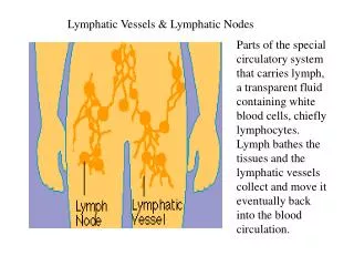
- The lymphatic system carries excess of the extracellular fluid back to the venous system. - This fluid is the result of filtration from capillaries. • - The lymphatic system consists of : • lymphatic vessels, • lymph nodes, • lymphatic ducts • Spleen.
Lymph vessels- The lymph capillaries start as blind ended vessels that collect to form lymphatic vessels which are similar to the small veins (contain smooth muscles) but contain valves so it has beaded appearance.- In the intestine the lymphatics are called lacteals.
Superficial lymphatic vessels: * Present on the deep surface of the epithelium. * They run parallel to the superficial blood vessels of the skin. * They then join together to form larger vessels, which pierce the deep fascia to join the deeper vessels.
Deep lymphatic vessels: * They are deep to the deep fascia, parallel to the major blood vessels. * These are larger than the superficial vessels and have thick walls and valves.
2. Lymph Nodes Definition: - They are small oval bodies along the course of lymphatic vessels. Site: - They form groups especially in the neck, axilla, thorax, abdomen, and groin.
Structure: - It has two surfaces: a. Convex (outer) surface: - This surface receives afferentlymphatics. • Filtrations of its contents occur inside the lymph node. b. Concave (inner) surface: - It is called the hilum. • This surface gives exit to an efferentlymphatic in addition to the presence of a small artery and vein.
Functions of lymph nodes: 1.Act as a filter as they prevent micro-organisms and certain substances from entering the blood stream. 2. Formation of lymphocytes. 3. Formation of antibodies. 4. In case of infection or malignancy, the lymph nodes become enlarged and change in consistency.
3. Lymphatic Duct a. Thoracic duct - It begins in the cisterna chili in the abdomen (in front of the lumbar vertebrae) - It ascends through the posterior abdominal and thoracic walls (deviating to the left side). - It terminates at the junction of left subclavian and left internal jugular veins. - It drains lymph from all the body exceptthe upper right quadrant. - There are two lymphatic ducts thoracic duct and right lymphatic duct.
b. Right lymphatic duct - It is much smaller in size. - It drains lymph from the upper right quadrant (right side of the head and neck, right upper limb, and right side of the chest) - It terminates at the junction of right subclavian and right internal jugular veins
Other lymphatic aggregations: They are seen in: - tonsils. - Mucous membrane of the intestine (Peyer's patches). - Spleen. - Thymus gland.
All parts of the body posses lymphaticsEXCEPT: • C.N.S: its lymphatics are replaced by perivascular spaces that are connected with the subarachnoid space. • Bone marrow. • Spleen. • Internal ear. • Epidermis. • Cartilage. • Bone.
On the other hand, certain parts of the body have a rich network of lymphatics e.g. • dermis of the skin, • mucous membranes, • serous membranes • glands.
Axillary Lymph Nodes Divided into 5 main groups: 1- Pectoral (Anterior) group 2- Scapular (Posterior) group 3- Lateral group 4- Infraclavicular LNs 5- Apical group
1- Pectoral (Anterior) group: Site: on lower border of pectoralis minor. Afferents: from front of upper ½ of trunk, and breast.
2- Scapular (Posterior) group: Site: along subscapular artery Afferents: 1- from the back of the upper ½ of the trunk, and 2- axillary tail of breast.
3- Lateral group: Site: Along the axillary vessels. Afferents: From upper limb.
4- Infraclavicular LNs: Site: Below clavicle Afferents: From the upper part of the breast.
5- Apical group: Site: At the apex of axilla Afferents: from previous groups. Efferents form the subclavian trunk which enters the thoracic duct or right lymph trunk
Applied note: Infection or malignancy in the upper limb or breast gives swelling in the axilla due to involvement of axillary LNs. • Virchow`s lymph node????
Inguinal lymph nodes I- Superficial inguinal LNs: Site: In the proximal region of the femoral triangle. Below the inguinal ligament No: 12- 20 Two groups: A- Horizontal group: i- Medial: It drains: Anterior abdominal wall below umbilicus, lower part of anal canal, external genitalia (male and female). ii- Lateral: It drains; buttock and back below iliac crest.
B- Vertical group: It drains the lower limb. It is present at the end of the great saphenous vein. All the above groups of LNs send efferents to deep inguinal and external iliac LNs.
Inguinal lymph nodes cont. II- Deep inguinal LNs: - Site: Deep to deep fascia on the medial side of the femoral vein. - No: 1-3. One of them may lie in the femoral canal (lymph node of Cloquet). - Afferent: from deep lymphatics of the lower limb and from superficial inguinal LNs. - Efferent: to external iliac LNs