Download
1 / 41
420 likes | 577 Vues
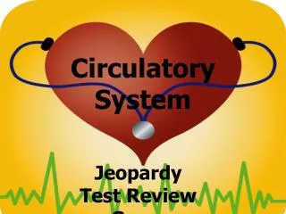
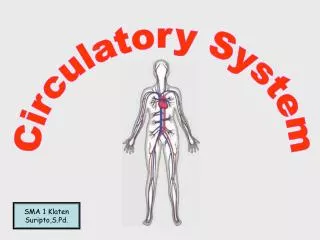
The Circulatory System – The Heart. Part 4: Regulation & Maintenance. The Heart. The Heart: The pump that supplies force critical to move blood through the circulatory system. Located in the thoracic cavity. 10 ounces total, 12 cm long, 9 cm wide.

E N D
The Circulatory System – The Heart Part 4: Regulation & Maintenance
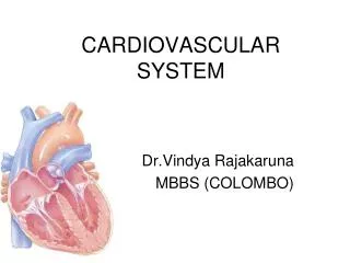
The Heart • The Heart: The pump that supplies force critical to move blood through the circulatory system. • Located in the thoracic cavity. • 10 ounces total, 12 cm long, 9 cm wide. • Great vessels attach at superior end, inferior end tapers off. • Mediastinum: Mass of tissue that extends from sternum to vertebral column between the lungs. Contains the heart!
The Heart • Pericardium: The double-walled membrane surrounding the heart. • Fibrous Pericardium: Outer membrane made of tough connective tissue providing protection & preventing overstretching of the muscle. • Serous Pericardium: Inner membrane. • Parietal Layer: Outer layer • Pericardial Cavity: Small gap that is filled with lubricating paricardial fluid. • Visceral Pericardium aka Epicardium: Inner layer
The Heart • Heart Wall: Made up of 3 layers! • Epicardium: The outer layer (and layer of the serous epicardium). • Composed of mesothelium & connective tissue. Provides smooth & slippery texture. • Myocardium: Middle layer. • Cardiac muscle that makes up the bulk of the heart & provide contraction force. • Endocardium: Smooth lining inside the chambers & valves; composed of endothelium.
The Heart • Chambers: 4 chambers total, 2 upper & 2 lower. • Atria: Upper chambers, both right & left. • Receive blood! • Auricle: A slight extension to each atria that increases the total volume. • Ventricles: Lower chambers, both right & left. • Pump blood out to the body. • Interatrial Septum: Separates the right & left atria. • Interventricular Septum: Separates the right & left ventricles.
The Heart • Valves: Control the one-way flow of blood through the chambers. 4 total: • Right Atrioventricular (A-V) Valve aka Tricuspid Valve: Regulates blood flow into the right ventricle. • Left Atrioventricular (A-V) Valve aka Bicuspid Valve aka Mitral Valve: Regulates blood flow into the left ventricle. • Aortic Valve: Regulates flow from the left ventricle to the aorta. • Pulmonary Valve aka Semi-lunar Valve: Regulates flow from the right ventricle to the pulmonary trunk. • Chordae Tendineae: Connects the Right & Left A-V Valves to the papillary muscles.
The Heart • Flow of Blood through the Heart: • Deoxygenated blood flows into the heart at the right atrium. • Blood passes from the right atrium to the right ventricle via the Right A-V valve. • Blood flows from the right ventricle through the pulmonary valve into the pulmonary trunk (and off to the lungs!). • Oxygenated blood re-enters the heart through the left atrium. • Blood passes from the left atrium through the left A-V valve to the left ventricle. • Blood flows from the left ventricle through the aortic valve into the aorta (and out to the body!).
The Heart • Flow of Blood Simplified: • Pulmonary Circuit: Right side of the heart pumps blood to the lungs only. • Systemic Circuit: Left side of the heart pumps blood to the rest of the body.
Disorders of the Heart Valves • Stenosis: Failure of a heart valve to fully open. • Insufficiency: Failure of a heart valve to fully close. • Mitral Valve Prolapse (MVP): Blood leaks back into the left atrium from the left ventricle.
Coronary Circulation • Left & Right Coronary Arteries: The first branches off the aorta that supply the left & right halves of the heart with blood. • Great & Middle Cardiac Veins: Return blood from the heart muscle itself to the coronary sinus & right atrium. • Anastomoses: The point where two or more branches of an artery serving the same area meet.
Cardiac Muscle Contraction • Cardiac Muscle: Striated, involuntary muscle specifically found in the heart. • Rhythmicity: Property of beating on a regular basis even without electrical stimulation. This is unique to cardiac muscle! • Myocytes: Ends of cardiac muscle cells joined together by intercalated discs. • Gap junctions between the discs allow for action potentials (for communication) & desmosomes (to hold the fibers together). • Endomysium: Surrounds the myocytes and supplies access to capillaries. 10X as many mitochondria as skeletal muscles.
Autorhythmic Conduction • Cardiac Conduction System: The electrical conduction system that triggers cardiac muscle contractions. • Composed of nodal tissue (special conductive tissue). • Sinoatrial (SA) Node: The pacemaker of the heart – sets the heart rhythm at appx. 72 beats per minute at rest. • Eptopic Pacemaker: Occurs when the heart rhythm is set by some other site than the SA node. • Arrhythmia: The term used for any abnormal cardiac rhythm.
Autorhythmic Conduction • Steps of Autorhythmic Conduction: • SA nodes send signals to the atrioventricular node. • Atrioventricular node sends signals through the atrioventricular bundle aka bundle of his to the ventricles. • Signal travels from the atrioventricular bundle to the remainder of the heart via Perkinje fibers.
Autorhythmic Conduction • Cardiovascular Center: Located in the medulla oblongata – receives sensory input from limbic system, cerebral cortex & sensory systems & interprets the need to decrease or increase heart rate. • Cardioacceleratory Center: Speeds the heart up. • Cardioinhibitory Center: Slow the heart down.
Measuring Cardiac Output • Electrocardiogram aka EKG: Records the electrical activity of the heart. • Electrocardiograph: The registered read out of the heart rhythm. Only shows the depolarization, not contraction of the muscle. • Three Deflections of the EKG: • P-Wave: Corresponds to the depolarization of the atria (Atrial contraction). • QRS-Wave: Corresponds to the depolarization of the ventricles (Ventricle contraction). • T-Wave: Corresponds to the ventricular repolarization.
Cardiac Cycle • Cardiac Cycle: Consists of one complete heartbeat. • Systole: The contraction phase. • Diastole: The relaxation phase.
Cardiac Cycle • 6 Phases of the Cardiac Cycle: • Rest: Atria are filling with blood; A-V valves open. • Atrial Systole: Atrial depolarization prompted by depolarization of the SA node. Forces blood into the ventricles (25 mL) over 0.1 seconds. • Ventricular Systole: Rising blood pressure in the ventricle triggers isovolumetric contraction, which leaves the volume the same but raises pressure. • Ventricular Ejection: When ventricular pressure is greater than aortal pressure, the semi-lunar valves open. • Stroke Volume: Total amount of blood moving into the pulmonary trunk & aorta (typically 70 mL). • Relaxation Period: Ventricular repolarization causes ventricular diastole – all 4 valves close for isovolumetric relaxation. • Ventricular Filling: A-V valve opens when ventricular pressure drops back below atrial pressure, allowing the ventricles to fill again. Lasts 250 msec.
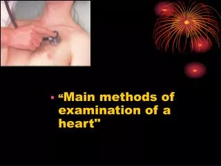
Heart Sounds • Heart Sounds: The sounds made by the opening & closing of the valves & the rush of blood. • First Heart Sounds (S1): The closing of the A-V valves, usually louder & longer lasting. • Second Heart Sounds (S2): The closing of the semi-lunar valves, usually quieter & quicker.
Heart Rhythm Disorders • Heart Murmer: Any abnormal clicking, gurgling, or rushing noise accompanying the heartbeat. • Tachycardia: Persistent resting heart rate of 100 BPM or higher. • Can be caused by stress, heart diseases, drugs, medications, or hyperthermia. • Bradycardia: Persistent resting heart rate of 60 BPM or less. • Can be caused by hypothermia; may be normal in athletes.
Cardiac Output • Cardiac Output: The amount of blood volume pumped by each ventricle per minute. • Cardiac Reserve: The difference between a resting cardiac output & the maximum potential cardiac output. • Heart Rate: The number of times the heart goes through one complete cardiac cycle per minute. • Adult males: 64-72 BPM resting. • Adult females: 72-80 BPM resting. • Infants: 120 BPM or more resting. • Cronotropic Agents: Anything that raises or lowers the rate of contraction. • Inotropic Agents: Alters the stroke volume of the ventricles. • Dopamine, epinephrine, & noreopinephine
Cardiac Output • Cardiac Output: Stroke volume X heart rate CO (mL/min). • SV (mL/beat) X HR (beats/min)
Chemicals Impacting Heart Rate • Sympathetic neurotransmitters & some hormones of the adrenal glands can accelerate the heart rate. • Hypernatremia: Excessive amounts of sodium - can lower the heart rate. • Hyperkalemia: Excessive amounts of potassium – can lower the heart rate and lead to death. • Hypercalcaemia: Excessive amounts of calcium – slows the heart rate. • Hypocalcaemia: Lowered amounts of calcium – speeds the heart rate up.
Stroke Volume • Stroke Volume: The amount of blood ejected by the ventricle during each contraction. Mostly determined by the amount of tension generated in the ventricles.
Stroke Volume • Regulated by 3 factors: • Preload: The greater the tension of the cardiac muscle prior to contraction, the greater the force of the contraction & the more blood that is expelled. Known as the Frank-Starling Law of the Heart. • Contractility: Strength of the contraction is enhanced by positive inotropic factors and decreased by negative inotropic agents. (Anything that stimulates or inhibits the sympathetic nervous system). • Afterload: The amount of pressure in the semi-lunar valves in the ventricles. The higher the pressure in the arteries, the less stroke volume.
Blood Pressure • Blood Pressure: The pressure in the heart and blood vessels. • The left side of the heart has higher blood pressure than the right, but both have the same volume of blood. • Pressure Gradient: The difference in pressure of the two sides of the heart & causes the valves to stay open. • Sphygmomanometer: The device used to measure the blood pressure.
Exercise & The Heart • Aerobic Exercise: Exercise that gets your heart pumping! • Can be good for the heart. • Increases cardiac reserve.