Download
1 / 32
360 likes | 827 Vues
Chapter 15 The Urinary System. Functions of the Urinary System. 1. Elimination of waste Nitrogenous wastes Toxins Drugs. 2. Regulates homeostasis Water balance Electrolytes Acid-base balance in the blood Blood pressure RBC blood cell production Activation of vit. D.

E N D
Functions of the Urinary System 1. Elimination of waste • Nitrogenous wastes • Toxins • Drugs 2. Regulates homeostasis • Water balance • Electrolytes • Acid-base balance in the blood • Blood pressure • RBC blood cell production • Activation of vit. D
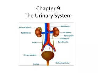
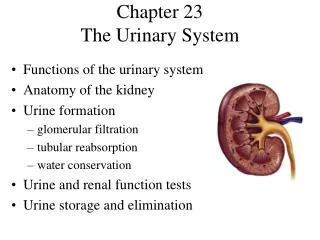
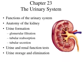
Organs of the Urinary system • Kidneys • Ureters • Urinary bladder • Urethra
Location of the Kidneys • Against dorsal wall • T12 to L3 • Right lower than left • Attached to ureters, renal blood vessels, & nerves at renal hilus • Atop kidney is adrenal gland
Coverings of the Kidneys 1. Renal capsule • Surrounds kidney 2. Adipose capsule • Surrounds kidney • protection • keeps kidney in location
Regions of the Kidney • Renal cortex – outer region • Renal medulla – inside the cortex • Renal pelvis – inner collecting tube
Kidney Structures • Medullary pyramids– triangular regions of tissue in medulla • Renal columns – extensions of cortex-like material inward • Calyces – cup-shaped structures that funnel urine towards renal pelvis
Nephrons • Structural and functional units of the kidneys • Forms urine • Main structures a. Glomerulus b. Renal tubule
Glomerulus • Specialized capillary bed • Arterioles on both sides (maintains high pressure) • Large afferent arteriole • Narrow efferent arteriole • Capillaries covered with podocytes from the renal tubule • Glomerulus sits within a glomerular capsule (1st part of the renal tubule)
Renal Tubule • Glomerular (Bowman’s) capsule • Proximal convoluted tubule • Loop of Henle • Distal convoluted tubule
Types of Nephrons 1. Cortical • entirely in cortex • most nephrons 2. Juxtamedullary • at boundary of cortex & medulla
Urine Formation Peritubular Capillaries • Arise from efferent arteriole • Normal, low pressure capillaries • Attached to venule • Cling to renal tubule • Reabsorb substances from collecting tubes • Filtration • Reabsorption • Secretion
Filtration • Nonselective passive process • Water and solutes smaller than proteins are forced through capillary walls • Blood cells cannot pass out to the capillaries • Filtrate is collected in the glomerular capsule and leaves via the renal tubule
Reabsorption • Peritubular capillaries reabsorb - Some water, Glucose, Amino acids, Ions • Some passive, most active • Most reabsorption occurs in proximal tubule Not Reabsorbed • Nitrogenous waste products - Urea - Uric acid - Creatinine • Excess water
Secretion – Reabsorption in Reverse • Materials move from peritubular capillaries into renal tubules • H+ & K+ • Creatinine • Materials left in renal tubule move to ureter
Formation of Urine Figure 15.5
Characteristics of Urine • Yellow due to pigment urochrome (from break-down of hemoglobin) & solutes • Sterile • Slightly aromatic • Normal pH of ~ 6 • Specific gravity of 1.001 to 1.035
Ureters • Slender tubes from kidney to bladder - Continuous with renal pelvis - Enter the posterior aspect of the bladder • Runs behind the peritoneum • Peristalsis aids gravity in urine transport
Urinary Bladder • Smooth, collapsible, muscular sac • Temporarily stores urine • Trigone – 3 openings - Two from ureters - One to urethrea
Urinary Bladder Wall • Detrusor muscle – pushes down • 3 layers of smooth muscle • Walls - thick & folded in empty bladder • Transitional epithelium - expands without increasing internal pressure
Urethra • Thin-walled tube • Carries urine from bladder by peristalsis • Release controlled by 2 sphincters • Internal urethral sphincter (involuntary) • External urethral sphincter (voluntary)
Urethra Gender Differences • Length - Females – 3–4 cm (1 inch) - Males – 20 cm (8 inches) • Location - Females – along wall of the vagina - Males – through the prostate and penis • Function - Females – only urine - Males –urine and sperm
Micturition (Voiding) Both sphincters must open • internal relaxes after bladder stretches • Activation - impulse to spinal cord and back via pelvic splanchnic nerves • external voluntarily relaxes
Maintaining Water Balance • Normal amount of water in humans - Adult females – 50% - Adult males – 60% - Babies – 75% - Old age – 45% • Water is necessary for many functions and levels must be maintained • Water intake = water output • Sources for water intake - foods and fluids - metabolic processes • Sources for water output - Vaporization from lungs - perspiration - feces - Urine
Distribution of Body Fluid • Intracellular fluid (inside cells) • Extracellular fluid (outside cells) - Interstitial fluid - Blood plasma Figure 15.7
Link Between Water and Salt • Changes in electrolyte balance causes water to move from one compartment to another - Alters blood volume & blood pressure - Can impair the activity of cells
Regulation of Reabsorption • Hormones - Antidiuretic hormone (ADH) prevents excessive water loss in urine • Aldosterone regulates sodium • Triggered by the renin-angiotensin mechanism • Monitored by kidneys and hypothalamus
Maintaining Blood pH • Normal Blood pH - Alkalosis – pH above 7.45 - Acidosis – pH below 7.35 • Most ions are metabolic byproducts • Most pH balance is maintained by kidneys • Other acid-base controlling systems - Blood buffers - Respiration
Blood Buffers • Molecules to prevent dramatic changes in [H+] - Bind to H+ when pH drops - Release H+ when pH rises • Three major chemical buffers • Bicarbonates • Phosphates • Proteins
Developmental Fetal/Newborn • Functional kidneys by 3rd month • Bladder is small in newborn • Urine cannot be concentrated • Control of voluntary sphincter ~ 18 mos • Urinary infections - common problems Aging • Bladder shrinks • Decline in function • Retention if prostate enlarges (males)