Download
1 / 35
360 likes | 829 Vues
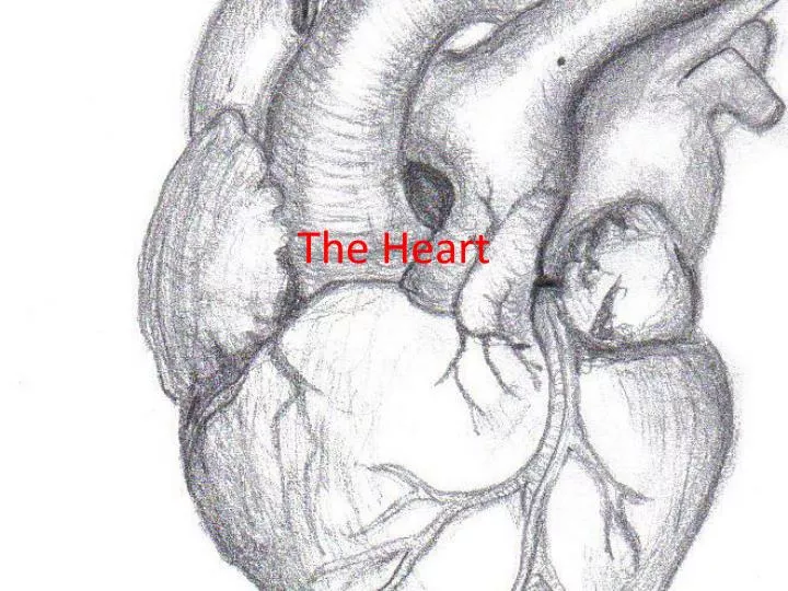
The Heart. OBJECTIVES. ANATOMY SHAPE POSITION STRUCTURE PHYSIOLOGY FLOW OF BLOOD THROUGH THE HEART BLOOD SUPPLY TO THE HEART CONDUCTING SYSTEM OF THE HEART CARDIAC OUTPUT . THE HEART. SHAPE ROUGHLY CONE SHAPED 10CM LONG SIZE OF THE OWNER’S FIST

E N D
OBJECTIVES • ANATOMY • SHAPE • POSITION • STRUCTURE • PHYSIOLOGY • FLOW OF BLOOD THROUGH THE HEART • BLOOD SUPPLY TO THE HEART • CONDUCTING SYSTEM OF THE HEART • CARDIAC OUTPUT
THE HEART • SHAPE • ROUGHLY CONE SHAPED • 10CM LONG • SIZE OF THE OWNER’S FIST • WEIGHS ABOUT 225g IN WOMEN AND ABOUT 310g IN MEN 10 CM CONE SHAPE
POSITION OF THE HEART • HEART LIES • IN THORACIC CAVITY IN MEDIASTINUM( i.e BETWEEN LUNGS) • OBLIQUELY, A LITTLE MORE LEFT TO LEFT THAN RIGHT • PRESENTS A BASE ABOVE AND APEX BELOW
POSITION OF THE HEART • ORGANS ASSOCIATED WITH THE HEART • INFERIORLY- DIAPHRAGM • SUPERIORLY- GREATER BLOOD VESSELS • LATERALLY- LUNGS • ANTERIORLY- STERNUM
STRUCTURE OF THE HEART • The heart wall • It is composed of three layers of tissue • PERICARDIUM • MYOCARDIUM • ENDOCARDIUM
PERICARDIUM • IT IS THE OUTERMOST LAYER, IT IS MADE UP OF 2 SACS • OUTER SAC- FIBROUS TISSUE, IT IS ADHERENT TO DIAPHRAGM( prevents overdistension of heart) • INNER SAC- SEROUS MEMBRANE(SM) • OUTER LAYER OF SM- PARIETAL PERICARDIUM • INNER LAYER OF SM-VISCERAL PERICARDIUM
MYOCARDIUM • COMPOSED OF CARDIAC MUSCLE • EACH FIBRE(CELL) HAS NUCLEUS AND ONE OR MORE BRANCHES • END OF CELL AND THEIR BRANCHES ARE IN CLOSE CONTACT- THESE JOINTS ARE CALLED AS INTERCALATED DISCS
ENDOCARDIUM • CONSISTS OF CHAMBERS AND VALVES OF THE HEART • THIN, SMOOTH, GLISTENING MEMBRANE • CONSISTS OF FLATTENED EPITHELIAL CELLS AND CONTINUOUS WITH ENDOTHELIAL LINING THE BLOOD VESSELS • PERMITS SMOOTH FLOW OF BLOOD INSIDE HEART
CHAMBERS OF THE HEART • DIVIDED INTO LEFT SIDE AND RIGHT SIDE BY SEPTUM
CHAMBERS OF THE HEARTsides are labeled in reference to the patient facing you • Two atria • Right atrium • Left atrium • Two ventricles • Right ventricle • Left ventricle --------------------------------------------------------------------------------
Valvesthree tricuspidone bicuspid (cusp means flap) • “Tricuspid” valve • RA to RV • Pulmonary or pulmonic valve • RV to pulmonary trunk (branches R and L) • Mitral valve (the bicuspid one) • LA to LV • Aortic valve • LV to aorta
FUNCTION OF SEMILUNAR VALVES (AORTIC AND PULMONIC VALVES)
THROUGH HEART SUPERIOR AND INFERIOR VENACAVAE EMPTY THEIR CONTENTS INTO RA BLOOD PASSES VIA AV VALVE TO RV AND IS THEN PUMPED TO PULMONERY ARTERY AND THEN TO LUNGS THROUGH PULMONIC VALVE TO LA VIA PULMONARY VEINS TO LV THROUGH MITRAL VALVE; TO BODY VIA AORTIC VALVE THEN AORTA
BLOOD SUPPLY TO THE HEART • ARTERIAL SUPPLY • SUPPLIED BY RIGHT AND LEFT CORONARY ARTERIES • VENOUS DRAINAGE • VENOUS BLOOD COLLECTED INTO A NUMBER OF CARDIAC VEINS , JOINS TO FORM CORONARY SINUS
CONDUCTING SYSTEM OF THE HEART • The heart pumps blood through the body • This is accomplished by contraction and relaxation of the cardiac muscle tissue in the myocardium layer. • Intercalated discs allow impulses to travel rapidly between adjacent cells so they function as one rather than individual cells
Conduction System Continued…. • Cardiac conduction system: The electrical conduction system controls the heart rate • This system creates the electrical impulses and sends them throughout the heart. These impulses make the heart contract and pump blood.
COMPONENTS OF THE CONDUCTION SYSTEM • Sino atrial Node (Part I): • located in back wall of the right atrium near the entrance of vena cava • initiates impulses 70-80 times per minute without any nerve stimulation from brain • establishes basic rhythm of the heartbeat • called the pacemaker of the heart • impulses move through atria causing the two atria to contract. • at the same time, impulses reach the second part of the conduction system
COMPONENTS OF THE CONDUCTION SYSTEM CONTINUED …. • Atrioventricular Node (Part II): • located in the bottom of the right atrium near the septum • cells in the AV node conduct impulses more slowly, so there is a delay as impulses travel through the node • this allows time for atria to finish contraction before ventricles begin contracting
ATRIOVENTRICULAR BUNDLE • A.K.A. “Bundle of His” • From the AV node, impulses travel through to the right and left bundle branches • These branches extend to the right and left sides of the septum and bottom of the heart.
ATRIOVENTRICULAR BUNDLE Continued…. • As the ventricles contract, blood is forced out through the semilunar valves into the pulmonary trunk and the aorta. • After the ventricles complete their contraction phase, they relax and the SA node initiates another impulse to start another cardiac cycle.
CARDIAC VOCABULARY • Contractility: Contractility is the intrinsic ability of cardiac muscle to develop force for a given muscle length. • Preload: Preload is the muscle length prior to contractility, and it is dependent of ventricular filling (or end diastolic volume…EDV) • This value is related to right atrial pressure. • The most important determining factor for preload is venous return.
Cardiac Vocabulary cont.. • Afterload: Afterload is the tension (or the arterial pressure) against which the ventricle must contract. • If arterial pressure increases, afterload also increases. • Afterload for the left ventricle is determined by aortic pressure • Afterload for the right ventricle is determined by pulmonary artery pressure.
CARDIAC OUTPUT Cardiac Output is the volume of blood pumped each minute, and is expressed by the following equation: • CO = SV x HR • Where: • CO is cardiac output expressed in L/min (normal ~5 L/min) • SV is stroke volume per beat • HR is the number of beats per minute