Download
1 / 14
220 likes | 837 Vues
Fertilization - fertilization in the upper third of the oviduct/fallopian tube - fertilization = union of egg and sperm to produce the zygote -sperm must undergo capacitation after ejaculation – increase rate of tail beating

E N D
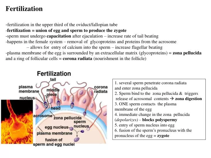
Fertilization -fertilization in the upper third of the oviduct/fallopian tube -fertilization = union of egg and sperm to produce the zygote -sperm must undergo capacitation after ejaculation – increase rate of tail beating -happens in the female system – removal of glycoproteins and proteins from the acrosome – allows for entry of calcium into the sperm – increase flagellar beating -plasma membrane of the egg is surrounded by an extracellular matrix (glycoproteins) = zona pellucida and a ring of follicular cells = corona radiata (nourishment in the follicle) 1. several sperm penetrate corona radiata and enter zona pellucida 2. Sperm bind to the zona pellucida & triggers release of acrosomal contents zona digestion 3. ONE sperm contacts the plasma membrane of the egg 4. immediate change in the zona pellucida (depolarizes) – blocks polyspermy 5. entry of sperm nucleus into egg 6. fusion of the sperm’s pronucleus with the pronucleus of the egg = zygote
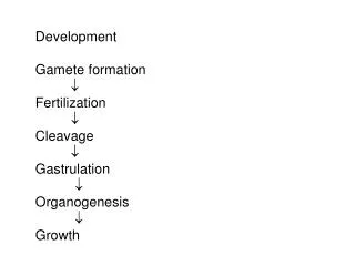
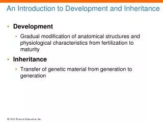
embryonic stage: 1st week to week 8 -first cell division – within 24 hrs & takes 6 hrs to complete -second day – four cells -end of third day – 16 cells -fourth day – morula stage -fourth to fifth day – blastocyst stage -end of fifth day – hatching of blastocyst from zona pellucida -6th day – implantation of blastocyst into enodmetrium • OVIDUCT: • -union of sperm and egg nuclei (zygote) -> first cell division within 24 hours to form • the embryo • cell division continues -> formation of the morulaat day 4 • morula = a mass of tiny, uniformly sized cells with equal amounts of cytoplasm • -cells of the embryo = blastomeres
embryonic stage: 1st week to week 8 -first cell division – within 24 hrs & takes 6 hrs to complete -second day – four cells -end of third day – 16 cells -fourth day – morula stage -fourth to fifth day – blastocyst stage -end of fifth day – hatching of blastocyst from zona pellucida -6th day – implantation of blastocyst into enodmetrium UTERUS: -day 5 – 6: morula forms a blastocyst = assymetrical ball of cells with a cavity -end of day 5 – blastocyst breaks out of the zona pellucida -day 6: -> implantation of blastocyst into the endometrium
-formation of the blastocyst marks the beginning of morphogenesis - shaping of the embryo, migration of dividing cells to specific locations -blastula = blastocyst - hollow ball of cells/blastomeres -outer layer = trophoblast - forms extraembryonic tissues (e.g. placenta, yolk sac) -inner mass of cells at one end - totipotent embryonic stem cells -second week of development (day 7 – 14) : -amniotic cavity forms between the inner cell mass and the trophoblast -the inner cell mass flattens = embryonic disc -disc – upper layer = epiblast & lower layer = hypoblast
-third week ( days 14 – 21): development of the gastrula via gastrulation (day 15) -gastrulation = conversion of the two-layered embryonic disk -> three embryonic germ layers by differentiation -differentiation - assumption of a specific cell structure and function -three germ layers: -> ectoderm (integumentary system & nervous system) -> mesoderm (mesenchyme bones, fat, cartilage, blood, muscle) -> endoderm (internal organs and membranes) -primitive streak: groove on the epiblast that forms before gastrulation -formation of the gastrula is identified through the formation of the primitive streak -cells pass from the epiblast through the primitive streak and form the mesoderm and endoderm during gastrulation through a process called invagination
-portions of the mesoderm that do not form the notochord segment into sections called somites -> specific body regions and structures • -four weeks into development (day 21 – 28): embryo forms a tubular structure & undergoes neurulation • -in front of the primitive streak is the primitive node • mesodermal cells from the primitive node become the notochord – forms at 22 days • neurulation: development of neural folds from ectoderm -> form into a neural tube • regions of the neural tube will form brain and spinal nerves, meninges • neural crest cells form skeletal and muscular components of the head • -17th day after fertilization – mesodermal cells form columns of mesoderm which segment into somites – these develop into skin, muscles and vertebrae in specific segments of the embryo
yolk sac chorion amnion placenta -yolk sac: forms blood cells, gives rise to sex cells and the stem cells of the immune system -also forms part of the embryonic digestive tube -portion of it becomes part of the umbilical cord -embryo’s first connection is via a connecting stalk -third week of development : formation of the allantois - tube from the yolk sac that projects into the connecting stalk - gives rise to the umbilical artery and vein
yolk sac placenta amnion chorion -chorion – forms after four weeks after implantation as slender projections that grow out from the trophoblast = chorionic villi -region of the chorion that is in contact with the endometrium becomes the placenta -produces human chorionic gondadotropin hormone – hormone of pregnancy. -amnion – surrounds the amniotic cavity (amnionic fluid + developing embryo) -amnion enfolds the connecting stalk and remnants of the yolk sac to form the umbilical cord
-fifth week (days 28 – 35): formation of lens, beginnings of maxilla and mandible & paddle-shaped forelimb -embryo is 10-12 mm long -35 + 2 days: formation of eye, ear, forebrain, nasal pit, tail -35 + 5 days: formation of midbrain, heart, external ear, primitive fingers -sixth week: formation of primitive toes (22-24 mm) -seventh week: formation of eyelids, webbed fingers -eighth week: separation of toes and fingers (34-40 mm)
-fetal stage : end of the eighth week -> birth -third month: body lengthens and head growth slows -ossification of bones -fourth month: reproductive organs appear -rapid body growth -lower limbs lengthen -development of hair, eyebrows, lashes, nipples -fifth month: growth slows -skeletal muscles are active -fetus curls into fetal position -sixth to ninth month: weight gain -skin smoothens as fat is deposited beneath skin -eyelids open -organs elaborate and grow (digestive and respiratory are last) -neuronal networks form
of every 100 oocytes: 69 implant 42 survive more than 1 week 37 survive more than 6 weeks 31 born alive
-umbilical vessels carry blood between the placenta and fetus -fetal blood - greater concentration of oxygen -enters fetus through umbilical vein and bypasses the fetal liver via the ductus venosus -enters the right atrium and can go to the right ventricle and out through the pulmonary artery (into the lungs) - bypasses the lungs through a vessel that connects to the aorta - ductus arteriosus -blood in the right atrium can directly flow into the left atrium (bypassing the lungs) through a hole between the atria called the foramen ovale -blood enters the left ventricle and exits via the aorta to travel to the rest of the body -exits the fetus via the umbilical artery (branch off the iliac artery)
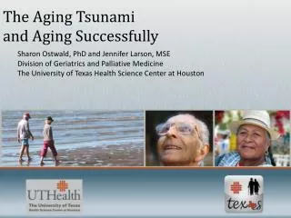
Aging Passive: breakdown of structures or failure of structures -connective tissue breaks down -DNA errors accumulate -free radical damage accumulates (free radical has an unpaired electron in its outer shell) Active: e.g. autoimmunity -begins before birth -certain cells undergo a programmed death = apoptosis -apoptosis starts in the embryo e.g. death of neurons as pathways are created Senescence: process of growing old -continuation of degeneration that begins during adulthood -result of normal wear and tear
Age-related changes: Integumentary system: Loss of collagen and elastic fibers, decrease of sweat and sebaceous glands, decrease in skin thickness, loss of hair pigments Skeletal system: loss of bone matrix, thinner, brittle bones, loss of height Muscular system: loss of muscle mass, degeneration of neuromuscular junctions Nervous system: loss of dendrites and synaptic connections, decrease in sensory sensitivity, decrease brain functions (memory, communication, smell & taste, loss of lens elasticity) Endocrine system: reduced hormone secretions, decreased metabolic rate Digestive system:decreased gastric motility, reduced gastric juice secretion CV system: degeneration of cardiac muscle, decrease in artery diameter