Download
1 / 34
450 likes | 893 Vues
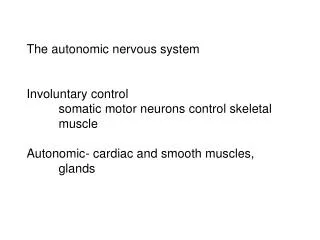
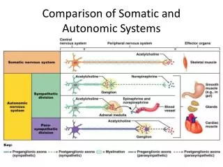
Comparison of Somatic and Autonomic Systems. Figure 14.2. Autonomic Nervous System (ANS). The ANS consists of motor neurons that: Innervate smooth and cardiac muscle and glands Make adjustments to ensure optimal support for body activities Operate via subconscious control

E N D
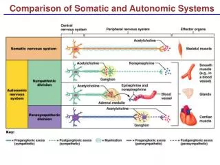
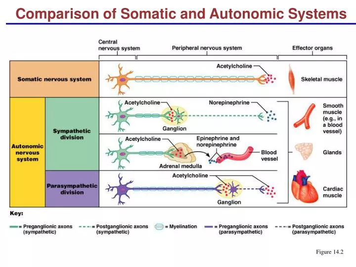
Comparison of Somatic and Autonomic Systems Figure 14.2
Autonomic Nervous System (ANS) • The ANS consists of motor neurons that: • Innervate smooth and cardiac muscle and glands • Make adjustments to ensure optimal support for body activities • Operate via subconscious control • Have viscera as most of their effectors
ANS Versus Somatic Nervous System (SNS) • The ANS differs from the SNS in the following three areas • Effectors • Efferent pathways • Target organ responses
Effectors • The effectors of the SNS are skeletal muscles • The effectors of the ANS are cardiac muscle, smooth muscle, and glands
Anatomy of ANS Figure 14.3
Efferent Pathways • Heavily myelinated axons of the somatic motor neurons extend from the CNS to the effector • Axons of the ANS are a two-neuron chain • The preganglionic (first) neuron has a lightly myelinated axon • The ganglionic (second) neuron extends to an effector organ
Neurotransmitter Effects • All somatic motor neurons release Acetylcholine (ACh), which has an excitatory effect • In the ANS: • Preganglionic fibers release ACh • Postganglionic fibers release norepinephrine or ACh and the effect is either stimulatory or inhibitory • ANS effect on the target organ is dependent upon the neurotransmitter released and the receptor type of the effector
Comparison of Somatic and Autonomic Systems Figure 14.2
Divisions of the ANS • The two divisions of the ANS are the sympathetic and parasympathetic • The sympathetic mobilizes the body during extreme situations • The parasympathetic performs maintenance activities and conserves body energy • The two divisions counterbalance each other’s activity
Role of the Parasympathetic Division • Concerned with keeping body energy use low • Involves the D activities – digestion, defecation, and diuresis • Its activity is illustrated in a person who relaxes after a meal • Blood pressure, heart rate, and respiratory rates are low • Gastrointestinal tract activity is high • The skin is warm and the pupils are constricted
Role of the Sympathetic Division • The sympathetic division is the “fight-or-flight” system • Involves E activities – exercise, excitement, emergency, and embarrassment • Promotes adjustments during exercise – blood flow to organs is reduced, flow to muscles is increased • Its activity is illustrated by a person who is threatened • Heart rate increases, and breathing is rapid and deep • The skin is cold and sweaty, and the pupils dilate
Comparison of Somatic and Autonomic Systems Figure 14.2
Anatomy of ANS Figure 14.3
Parasympathetic Division Outflow Figure 14.4
Sympathetic Outflow • Arises from spinal cord segments T1 through L2 • Sympathetic neurons produce the lateral horns of the spinal cord • Preganglionic fibers pass through the white rami communicantes and synapse in the chain (paravertebral) ganglia • Fibers from T5-L2 form splanchnic nerves and synapse with collateral ganglia • Postganglionic fibers innervate the numerous organs of the body
Sympathetic Outflow Figure 14.5
Neurotransmitters and Receptors • Acetylcholine (ACh) and norepinephrine (NE) are the two major neurotransmitters of the ANS • ACH is released by all preganglionic axons and all parasympathetic postganglionic axons • Cholinergic fibers – ACH-releasing fibers • Adrenergic fibers – sympathetic postganglionic axons that release NE or E • Neurotransmitter effects can be excitatory or inhibitory depending upon the receptor type
Cholinergic Receptors • The two types of receptors that bind ACh are nicotinic and muscarinic • These are named after drugs that bind to them and mimic ACh effects
Nicotinic Receptors • Nicotinic receptors are found on: • Motor end plates (somatic targets) • All ganglionic neurons of both sympathetic and parasympathetic divisions • The hormone-producing cells of the adrenal medulla • The effect of ACh binding to nicotinic receptors is always stimulatory
Muscarinic Receptors • Muscarinic receptors occur on all effector cells stimulated by postganglionic cholinergic fibers • The effect of ACH binding: • Can be either inhibitory or excitatory • Depends on the number of receptor type of the target organ
Adrenergic Receptors • The two types of adrenergic receptors are alpha and beta • Each type has two or three subclasses (1, 2, 1, 2 , 3) • Effects of NE binding to: • receptors is generally stimulatory • receptors is generally inhibitory • A notable exception – NE binding to receptors of the heart is stimulatory
Effects of Drugs • Atropine – blocks parasympathetic effects • Neostigmine – inhibits acetylcholinesterase and is used to treat myasthenia gravis • Tricyclic antidepressants – prolong the activity of NE on postsynaptic membranes • Over-the-counter drugs for colds, allergies, and nasal congestion – stimulate -adrenergic receptors • Beta-blockers – attach mainly to 1 receptors and reduce heart rate and prevent arrhythmias
Drugs that Influence the ANS Table 14.4.1
Drugs that Influence the ANS Table 14.4.2
Interactions of the Autonomic Divisions • Most visceral organs are innervated by both sympathetic and parasympathetic fibers • This results in dynamic antagonisms that precisely control visceral activity • Sympathetic fibers increase heart and respiratory rates, and inhibit digestion and elimination • Parasympathetic fibers decrease heart and respiratory rates, and allow for digestion and the discarding of wastes
Sympathetic Tone • The sympathetic division controls blood pressure and keeps the blood vessels in a continual state of partial constriction • This sympathetic tone (vasomotor tone): • Constricts blood vessels and causes blood pressure to rise as needed • Prompts vessels to dilate if blood pressure is to be decreased • Alpha-blocker drugs interfere with vasomotor fibers and are used to treat hypertension
Parasympathetic Tone • Parasympathetic tone: • Slows the heart • Dictates normal activity levels of the digestive and urinary systems • The sympathetic division can override these effects during times of stress • Drugs that block parasympathetic responses increase heart rate and block fecal and urinary retention
Cooperative Effects • ANS cooperation is best seen in control of the external genitalia • Parasympathetic fibers cause vasodilation and are responsible for erection of the penis and clitoris • Sympathetic fibers cause ejaculation of semen in males and reflex peristalsis in females
Unique Roles of the Sympathetic Division • Regulates many functions not subject to parasympathetic influence • These include the activity of the adrenal medulla, sweat glands, arrector pili muscles, kidneys, and most blood vessels • The sympathetic division controls: • Thermoregulatory responses to heat • Release of renin from the kidneys • Metabolic effects
Thermoregulatory Responses to Heat • Applying heat to the skin causes reflex dilation of blood vessels • Systemic body temperature elevation results in widespread dilation of blood vessels • This dilation brings warm blood to the surface and activates sweat glands to cool the body • When temperature falls, blood vessels constrict and blood is retained in deeper vital organs
Levels of ANS Control • The hypothalamus is the main integration center of ANS activity • Subconscious cerebral input via limbic lobe connections influences hypothalamic function • Other controls come from the cerebral cortex, the reticular formation, and the spinal cord
Levels of ANS Control Figure 14.9