Download
1 / 25
270 likes | 530 Vues
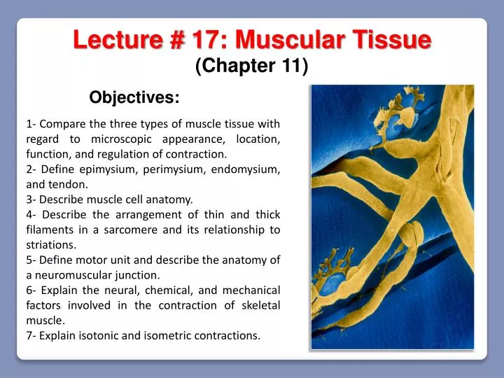
Lecture # 17: Muscular Tissue. (Chapter 11). Objectives:. 1- Compare the three types of muscle tissue with regard to microscopic appearance , location, function, and regulation of contraction. 2- Define epimysium , perimysium, endomysium , and tendon.

E N D
Lecture # 17: Muscular Tissue (Chapter 11) Objectives: 1- Compare the three types of muscle tissue with regard to microscopic appearance, location, function, and regulation of contraction. 2- Define epimysium, perimysium, endomysium, and tendon. 3- Describe muscle cell anatomy. 4- Describe the arrangement of thin and thick filaments in a sarcomere and its relationship to striations. 5- Define motor unit and describe the anatomy of a neuromuscular junction. 6- Explain the neural, chemical, and mechanical factors involved in the contraction of skeletal muscle. 7- Explain isotonic and isometric contractions.
Skeletal Muscle (attachments between muscle and bone matrix) Tendon Muscle Fascicle (connective tissue surrounding entire muscle) Muscle fiber (cell) Epimysium (connective tissue around muscle cells) Endomysium (connective tissue around muscle fascicles) Perimysium
The Muscle Fiber Nucleus T tubules Muscle fiber They conduct the nerve impulse from the sarcolemma to the interior of the cell Terminal cisterna Sarcoplasmicreticulum It stores and releases calcium for muscle contraction Mitochondria They produce the chemical energy (ATP) for muscle contraction Openings intotransverse tubules Triad: 2 Terminal cisternae Myofibrils Transverse tubule Sarcolemma Sarcoplasm Myofilaments
SKELETAL MUSCLE Contains: Surrounded by: Epimysium Muscle fascicles MUSCLE FASCICLE Contains: Surrounded by: Perimysium Muscle fibers (cells) MUSCLE FIBER (CELL) Contains: Surrounded by: Endomysium Myofibrils MYOFIBRIL Contains: Myofilaments They are organized in sarcomeres MYOFILAMENTS Thick filaments: myosin Thin filaments: actin Sarcomere
Myofilaments Thick filament Thin filament G actin Tail Myosin molecule Head Hinge region Contractile proteins Regulatory proteins Tropomyosin Troponin complex They do the work of shortening the muscle fiber They act like a switch to determine when the fibers can contract Troponin Myosin Tropomyosin Actin
Striations They attach the thin and elastic filaments Titin (elastic filaments) Sarcomere They are the smallest functional units of the muscle fiber M line Z line Z line I band(lighter) It contains thin filaments but not thick filaments Zone of overlap H band Zone of overlap Myosin (thick filaments) Actin (thin filaments) A band (dark) M line: It consists of proteins that connect each thick filament with its neighbors A band: Its length is equal to the length of the thick filaments. It contains both thin and thick filaments H band: It is a lighter region on either side of the M line, which contains only thick filaments I band Z line Zone of overlap Zone of overlap:It is the region where the thin filaments are situated between the thick filaments H band M line
A band H band I band I band Zone of overlap M line Z line Z line Zone of overlap When a skeletal muscle fiber contracts: 1- The H bands and I bands get smaller 3- The Z lines move closer together 2- The zone of overlap get larger 4- The width of the A band remain constant
ATP Contraction ADP Pi ADP Pi ADP Pi Active sites Hydrolysis of ATP to ADP + Pi; activation and cocking of myosin head Formation of myosin–actin cross-bridge Sliding of thin filament over thick filament shorten the sarcomeres and muscle also shorten (contraction) Power stroke; sliding of thin filament over thick filament
ATP Hydrolysis of ATP to ADP + Pi; activation and cocking of myosin head Binding site for myosin (active site) G-actin strand ADP + Pi
+2 At low intracellular concentration of Ca the tropomyosin blocks the binding sites for myosin in the actin molecules and prevents the formation of cross-bridges Troponin Ca +2 F-actin strand Tropomyosin Cross-bridge ADP + Pi
+2 At high intracellular concentration of Ca the troponin is activated and undergoes a conformational change that moves the tropomyosin away from actin’s binding sites for myosin heads Troponin Ca Ca Ca Ca Ca Ca Ca Ca Ca Ca Ca Ca Ca Ca +2 +2 +2 +2 +2 +2 +2 +2 +2 +2 +2 +2 +2 +2 F-actin strand Tropomyosin Cross-bridge ADP + Pi
The Nerve-Muscle Relationship Motor unit: It is one nerve fiber and all the muscle fibers innervated by it The average motor unit contains 200 muscle fibers for each motor unit Neuromuscular junction (NMJ): It is the point where a nerve fiber meets a muscle fiber
Action potential The Muscle Fiber Nucleus Action potential Synaptic knob or axon terminal T tubules Ca2+ Ca2+ Ca2+ Ca2+ Ca2+ Ca2+ Muscle fiber They conduct the nerve impulse from the sarcolemma to the interior of the cell. Terminal cisterna Sarcoplasmicreticulum It stores and releases calcium for muscle contraction. Mitochondria They produce the chemical energy (ATP) for muscle contraction. . . .. . . .. . ... Neurotransmitter Synaptic cleft Myofibrils Myofilaments
The Neuromuscular Junction Myelin Motor nerve fiber Synaptic knob Synaptic knob Sarcolemma Mitochondrion Sarcolemma T tubule • They have ACh receptors which bind Ach Junctional folds Synaptic cleft Sarcoplasm Synaptic vesicle • They contain acetylcholine (Ach) Myofilaments
Two Types of Ion Channels Ligands (Neurotransmitters, hormones) + + + + + + + _ _ _ _ _ _ _ _ _ _ _ + + + + + Chemically gated ion channels: Voltage gated ion channels: They open when the specific ligand binds to the receptor. They open in response to a voltage change in the plasma membrane.
Chemically Gated ion Channels Acetylcholine Na+ _ _ _ + + + Resting Membrane Potential End plate Potential _ _ _ + + +
Excitation Action potential Axon of motor neuron 1- The Arrival of an Action Potential 2- The Release of Acetylcholine Synaptic terminal Sarcolemma Action potential 2+ Mitochondrion Ca Voltage gated ion channels open Sarcolemma T tubule Junctional folds Synaptic cleft Synaptic vesicle Fusing synaptic vesicle
Axon terminal 3- Binding of Ach to the receptors 4- Opening of ligand-gated ion channels and creation of end plate potential + + + + + + + + _ _ _ _ _ _ _ _ _ _ _ Motor End Plate Acetylcholine End-plate potential Chemically gated ion channels + It is rapid fluctuation in the membrane potential that falls back to a level close to the resting membrane potential Na + K Acetic acid Choline
5- Opening of voltage-gated ion channels and creation of action potential Action Potential: It is a rapid voltage change in which a plasma membrane reverses its electrical polarity Action potential have self-propagating effect that produce a traveling wave of excitation in the nerves and muscles cells + + + _ _ _ _ + + + + + _ _ _ _ _ _ _ _ + + + + End-plate potential Voltage-gated ion channels Action Potential Ligand- gated ion channels
ATP 1 & 2- Nerve signal stimulates voltage-gated calcium channels that result in exocytosis of synaptic vesicles containing ACh = ACh release +2 Ca Acetylcholine + Na - - + + + + + + + + + + + + - - - - - - - - - - - - 5- Voltage change in end-plate region (EPP) opens nearby voltage-gated channels in plasma membrane producing an action potential 6 & 7- Action potential spreading over sarcolemma reaches and enters the T tubules -- voltage-gated channels open in T tubules causing calcium gates to open in SR 3 & 4- Binding of ACh to the surface of muscle cells opens Na+ and K+ channels resulting in an end-plate potential (EPP) 12 & 13- Nerve stimulation ceases and acetylcholinesterase removes ACh from receptors so stimulation of the muscle cell ceases 8 & 9- Calcium released by SR binds to troponin. Troponin-tropomyosin complex changes shape and exposes active sites on actin 14- Active transport pumps calcium from sarcoplasm back into SR where it binds to calsequestrin. ATP is needed for muscle relaxation as well as muscle contraction 10 - Myosin ATPase in myosin head hydrolyzes an ATP molecule, activating the head and “cocking” it in an extended position. It binds to an active site on actin 11 – Myosin releases the ATP and P and flexes into a bent , tugging the thin filaments along with it (POWER STROKE). 15 & 16- Loss of calcium from sarcoplasm results in troponin-tropomyosin complex moving over the active sites which stops the production or maintenance of tension Active transport Muscle fiber returns to its resting length due to stretching of series-elastic components and contraction of antagonistic muscles
Rigor Mortis • Stiffening of the body beginning 3 to 4 hours after death -- peaks at 12 hours after death & diminishes over next 48 to 60 hours • Deteriorating sarcoplasmic reticulum releases calcium • Activates myosin-actin cross bridging & muscle contracts, but does not relax. • Muscle relaxation requires ATP & ATP production is no longer produced after death • Fibers remain contracted until myofilaments decay