Download
1 / 6
60 likes | 187 Vues
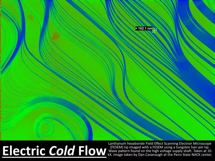
Lanthanum hexaboride Field Effect Scanning Electron Microscope (FESEM) tip imaged with a FESEM using a tungsten hair-pin tip . Wave pattern found on the high voltage supply shaft. Taken at 10 kX . Image taken by Dan Cavanaugh at the Penn State NACK center. Electric Cold Flow.

E N D
Lanthanum hexaborideField Effect Scanning Electron Microscope (FESEM) tip imaged with a FESEM using a tungsten hair-pin tip. Wave pattern found on the high voltage supply shaft. Taken at 10 kX. Image taken by Dan Cavanaugh at the Penn State NACK center. Electric Cold Flow
Field Effect Scanning Electron Microscope (FESEM) image of zinc oxide nano-shell. ZnO2 nano-shells grown by pyrolysis in an Low Pressure Chemical Deposition (LPCVD). Center particle is 10 µm dia. Image taken by Dan Cavanaugh at the Penn State NACK center. My Pet “Spike”
Field Effect Scanning Electron Microscope (FESEM) image of resist patterned by nano-imprinting. Base widths are approximately 200 nm. False-color added with ImageJ freeware. Image taken by Suxing Pan at the Penn State NACK center. Infinite Isosceles Integration
Field Effect Scanning Electron Microscope (FESEM) image of carbon nano-wires (magenta-navy blue) with iron cap (orange). The nano-wires were formed by vapor-liquid-solid (VLS) growth in an Low Pressure Chemical Deposition (LPCVD). Wires are approximately 100 nm dia. by several microns long. Image taken by Dan Cavanaugh at the Penn State NACK center. Sprouts of the Future
Field Effect Scanning Electron Microscope (FESEM) image of zinc oxide nano-wires. ZnO2 nano-wires grown by pyrolysis in an Low Pressure Chemical Deposition (LPCVD). Hundred’s of nano-wires (approximately 50 nm dia.) splitting from precursor nodes (approximately 10 µm wide) . Image taken by Dan Cavanaugh at the Penn State NACK center. Nano-Thistle
Field Effect Scanning Electron Microscope (FESEM) image of zinc oxide nodes (precursors for nano-wire). ZnO2 nodes grown by pyrolysis in an Low Pressure Chemical Deposition (LPCVD). Image taken by Dan Cavanaugh at the Penn State NACK center. Nano-Broccoli