Download
1 / 2
20 likes | 151 Vues
以 Porphyromonas gingivalis 之脂多醣和白介質 -1b 刺激受尼菲迪平誘導增生牙齦細胞觀察男性荷爾蒙接受體、白介質 -6 與 Th1 / Th2 細胞激素之表現.

E N D
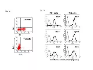
以Porphyromonas gingivalis之脂多醣和白介質-1b刺激受尼菲迪平誘導增生牙齦細胞觀察男性荷爾蒙接受體、白介質-6與Th1 / Th2細胞激素之表現 我們於2001年針對因尼菲迪平(nifedipine)引發牙齦増生患者之増生牙齦(nifedipine induced gingival overgrowth, NIGO)的組織以及其他的牙周組織進行Th1 / Th2的免疫染色,結果在NIGO病人的牙齦中,可以看到明顯的男性荷爾蒙接受體(androgen receptor, AR)與Th1的表現,在此in vivo的研究我們提出AR的增強與Th1 cytokine的表現可能是導致NIGO致病機轉的假說。 因此本實驗延續上述in vivo的臨床實驗,我們在牙周手術區域取下健康H組(n=4, age: 30-39 y/o)和NIGO組(n=4, age: 48-65 y/o)的牙齦纖維母細胞,在體外培養到第三代後,將牙周致病菌Porphyromonas gingivalis萃取出之脂多醣內毒素(lipopolysaccharide, LPS),以濃度10 μg/ml和IL-1b 10 ng/ml刺激48小時後,以不加任何藥物作為控制組。其後分離上清液來做酵素連結免疫吸附分析(enzyme-linked immunosorbent assay, ELISA),以測量細胞激素IL-2、IL-4、IL-6的表現,而細胞層則透過聚合酶連鎖反應(polymerase chain reaction, PCR)探測IL-2、IL-4、IL-6和AR的cDNA基因表現。結果發現在IL-2和IL-4不論是在上清液或是細胞層的表現都低於可偵測值,而AR和IL-6則在cDNA和ELISA的表現都有可偵測的增加。我們將PCR產物在紫外光下連接Gel Doc的應用程式做半定量,得到之數值以Mann-Whitney U Test做組間的統計,發現這兩種(H、NIGO)細胞在AR和IL-6的表現上,經過IL-1b刺激48小時後,都有顯著的增加,其p<0.05;而經過LPS刺激之組別與控制組則在AR和IL-6表現上,並沒有統計上之差異;而IL-6的ELISA表現以相同統計方式所得結果亦是無統計上之差異。另外若以Spearman Rank Correlation Coefficient來統計IL-6和AR的相關性,則發現不論是何種藥物刺激,彼此之間並無顯著的相關。 牙周病致病機轉除了細菌之外,其所引發之炎性反應,也會引發許多細胞激素的參予。因此我們透過上述實驗可知,炎前細胞激素(IL-1b)對NIGO細胞所引起的反應也較細菌內毒素的影響為大,而NIGO的細胞對於IL-1b的刺激感受性亦強於正常的牙齦纖維母細胞。因此對於NIGO的病人,我們除了以傳統的牙周治療以控制牙周炎性反應之外,也可併用抗發炎用藥或生物製劑,以減緩因牙周發炎而引起NIGO之牙齦腫大現象。
Expression of androgen receptor, interleukin-6, and Th1 / Th2 cytokines in the cells derived from nifedipine induced gingival overgrowth tissue stimulated with Porphyromonas gingivalis lipopolysaccharide and interleukin-1b • 。From immunostaining results of Th1/Th2 of nifedipine induced gingival overgrowth(NIGO)tissues and other periodontal tissues in 2001, a significant expression of androgen receptor(AR)could be observed. Using these results in vivo, we addressed the hypothesis that AR’s improvement and Th1 cytokine’s expression might be the pathogenic factors to NIGO. The present study extended above clinical trials in vivo. Gingival fibroblast from healthy, H(n=4, age: 30-39 y/o)tissues and NIGO(n=4, age: 48-65 y/o)tissues were handled from periodontal surgery area and put into cell culture. After third generation, pre-determined IL-1β and LPS of Porphyromonas gingivalis were used to stimulated sample cells for 48 hours. The concentration of the pre-determined extract was 10 μg/ml for LPS endotoxins and 10 ng/ml for IL-1β, respectively. These gingival fibroblasts were used as our control group in this experiment. Expression of IL-2、IL-4 and IL-6 extracted from supernatant were measured by ELISA. On the other hand, Cell layer extracted from pellet were analyzed by PCR to detect expression level of mRNA of IL-2、IL-4、IL-6 and AR. After 48 hours induction, the expression of IL-2 and IL-4 is under detetable value in this study either in supernatant or cell layer. Increased expression was detectable in the part of AR and IL-6 through mRNA level and ELISA. We used Gel Doc software to semi-quantify our PCR product under ultraviolet and studied the statistic difference between groups by Mann-Whitney U software. Results with p<0.05 showed that the amount of AR and IL-6 form these two groups(H and NIGO)were increased after IL-1β induction for 48 hours. However, the difference of AR and IL-6 between control group and experimental group with LPS stimulated was not remarkable. The information from using Spearman Rank Correlation Coeffienct for IL-6 and AR studies also showed no significant results while stimulated by different drugs. Besides bacteria, pathogenic mechanism of periodontal disease could not only induced inflammation but also evoke lots of cytokines to participate in. Through above experiments, we discovered NIGO cells were more sensitive to the induction of IL-1b than by P. gingivalis - LPS. It implies that in the treatment of NIGO, traditional periodontal treatment adjusted with anti-inflammatory drugs and biological agents can be conducted to inhibit both the effects of periodontal pathogen and pre-inflammatory cytokine on NIGO gingival fibroblast