Download
1 / 67
670 likes | 678 Vues
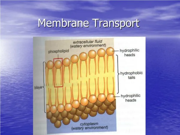
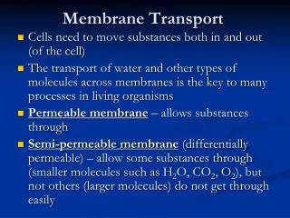
Membrane Transport. ECB Ch 12 (the passage of small water-soluble molecules through the cell membrane). The interior of the lipid bilayer is hydrophobic, the plasma membrane tends to block the passage of almost all water-soluble molecules.

E N D
Membrane Transport ECB Ch 12 (the passage of small water-soluble molecules through the cell membrane)
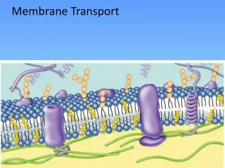
The interior of the lipid bilayer is hydrophobic, the plasma membrane tends to block the passage of almost all water-soluble molecules. • The membrane transport proteins: span the membrane, providing private passageways across the bilayer for select substance (small water-soluble molecules). 12_01_transport_prot.jpg • each type of transport protein in a cell membrane transfers a particular type of molecule, causing a selective set of solutes to end up inside the membrane-enclosed compartment.
The ion concentrations inside a cell are very different from those outside • Ions are crucial for a cell’s survival and function. • Ions in a cell’s environment and their movements across cell membranes play an essential part in many biological processes.
simple diffusion 12_02_diffusion_rate.jpg (nonpolar) (size, solubility properties) • The interior of the lipid bilayer is hydrophobic, the plasma membrane tends to block the passage of almost all water-soluble molecules. • Lipid bilayers are impermeable to solutes and ions.
Two main classes of membrane transport proteins • carrier proteins • which have moving parts, can shift small molecules from one side of the membrane to the other by changing their shape • transported solute: small organic molecules or inorganic ions • channel proteins • from tiny hydrophilic pores in the membrane through which solutes can pass by diffusion • most channel proteins let through inorganic ions only (therefore called ion channels) The membrane transport proteins that have been studied in detail shows the structure of multipass transmembrane proteins.
12_03_carrier_channel.jpg A carrier protein allow passage only to solute molecules that fit into a binding site on the protein. A channel protein discriminates mainly on the basis of size and electric charge. • Channel proteins transport molecules at a much greater rate than carrier proteins. • Channel opening and closing is usually controlled by an external stimulus or by conditions within the cell.
12_04_pass_act_transport.jpg (facilitate diffusion) (All channel proteins and many carrier proteins act such a way) (carrier proteins only)
Carrier proteins are required for transport of almost all small organic molecules across cell membranes. • Each cell membrane has its own characteristic set of carrier proteins. 12_05_carrier_proteins.jpg
To understand fully how a carrier protein transfers solutes across a membrane, we would need to know it’s 3-D structure in detail. • bacteriorhodopsin: a light-activated H+ pump • Ca2+ pump: moves Ca2+ from the cytosol into the sarcoplasmic reticulum sarcoplasmic reticulum: a specialized form of endoplasmic reticulum found in skeletal muscle cells
bacteriorhodopsin 11_28_Bacteriorhodop.jpg seven α helices polar a.a. side chain • generating a [H+]gradient (energy store) • driving ATP synthase (a membrane protein) generating ATP • retinal: • a light-absorbing non-protein molecule • deep purple color
recover from the contraction 12_06_Ca_pump.jpg stimulate cell contracts Ca2+ Ca2+ lead through the protein, allowing the ion to avoid contact with the lipid bilayer
passive transport of a carrier protein -by concentration gradient 12_07_conforma_change.jpg conformational change ex: glucose carrier Highly selective: bind only D-glucose
12_08_electroch_gradient .jpg ex: Na+ ex: K+: The cytoplasmic side of the plasma membrane is usually at a negative potential. The [K+] is higher inside cells than out side. There is little net movement of K+ across the membrane.
12_09_active_transport.jpg Cells carry out active transport in three main ways:
The Na+-K+ ATPase (The Na+-K+ pump) 12_10_Na_K_pump.jpg
phosphorylation 12_12_Na_K_cyclic.jpg (takes ~10 millisecond) dephosphorylation The Na+-K+ pump transports ions in a cyclic manner.
12_13_Carrier_proteins.jpg the same direction opposite directions ex: glucose-Na+ symports, Na+-H+ exchanger: a Na+-driven antiport
glucose-Na+ symport 12_14_symport.jpg • using the electrochemical Na+ gradient to drive the import of glacose • binding of Na+ inducing a conformational change in the protein that greatly increasing the protein’s affinity for glucose
12_15_glucose_gut.jpg uniport The two types of glucose carriers are kept segregated in their proper domains of the plasma membrane by a diffusion barrier formed by a tight junction.
Other Na+-transporters • Cells in the lining of the gut and in many other organs contain a variety of symports in their plasma membrane that are similarly driven by the electrochemical gradient of Na+ each of themspecifically imports sugars, amino acids, …… into the cell. • Na+-driven antiports are also important, such as Na+-H+ exchanger in the plasma membranes of many animal cells, is one of the main devices that cells use to control their pH in their cytosol.
osmosis • osmotic pressure • the Na+-K+ pump helps maintain the osmotic balance of animal cells
12_17_osmotic_swelling.jpg Na+-K+ pump cell wall contractile vacuoles turgor pressure
Ca2+ pump • drive by ATP (an ATPase) • maintain the low concentration of Ca2+ in the cytosol • Ca2+ is often used as a signal to trigger other intracellular events • the lower the background concentration of free Ca2+ the more sensitive the cell is to an increase in cytosolic Ca2+ • the eucaryotic cells in general matain very low concentrations of free Ca2+ in their cytosol (~10-4 mM) in the face of very much higher extracellular Ca2+ concentrations (~1-2 mM) • it is archived mainly by means of Ca2+ pump in both the plasma membrane and the ER membrane, which actively pump Ca2+ out of the cytosol
ATP-driven pumps (Na+-K+ pumps, Ca2+ pumps,……) have similar amino acid sequences and structures • with about 10membrane-spanning α helices in each subunit • it is likely that they have a common evolutionary origion
The H+ pumps • Plant cells, fungi (including yeasts), and bacteria do not have Na+-K+ pumps in their plasma membrane (instead of an electrochemical gradient of Na+) • They rely on the electrochemical gradient of H+ to drive the transport of solute into the cell • The H+ gradient is created by H+ pumps in the plasma membrane • The H+ pump also creates an acid pH in the medium surrounding the cell • In some photosynthetic bacteria the H+ gradient is created by the light-driven H+ pumps such as bacteriorhodopsin • Plant, fungi and many other bacteria, their H+ pumps are driven by ATP (as the ATPase)
A different type of H+ ATPase is found in in the membranes of some intracellular organells such as lysomes (animal cells) and central vacuole (plant and fungal cells). • Their function is to pump H+ out of the cytosol into the organelle, helping to keep the pH of the cytosol neutral and the pH of the interior of organelle acidic.
Channel proteins • A few channel proteins form relatively large pores • gap junctions (channels between two adjacent cells) • porins (channels in the outer membrane of mitochondria and some bacteria)
21_28_Gap_junctions.jpg The gap junctions
11_25_Porin.proteins.jpg The porin protein
Ion channels • Almost all of the channel proteins in the plasma membrane of animal and plant cella are ion channels. • narrow, highly selective pores concerned exclusively with the transport of inorganic ions, mainly Na+, K+, Cl-, Ca2+ • Distinguish from simple aqueous pore: • ion selectivity • depends on the diameter, shape of the ion channel and on the distribution of charged a. a. in its lining • gated (not continuously open) • Advantages over carrier proteins • With a transport rate (more than a million ions can pass through one channel each second) 1000 times greater than the fast known carrier protein
K+ channel 12_19_selectivity_filter.jpg bear a partial negative charge and form transient binding site for the K+ that have shed their watery shells
12_20_ion channel.jpg Most ion channels are not continuously open, they are gated.
membrane potential: the voltage across the membrane • When a ion channel opens, ions rush through it that changes the membrane potential, thus forcing other membrane potential sensitive ion channels to open or close in a matter of milliseconds. • The resulting flurry of electrical activity can spread rapidly from one region of the cell membrane to another conveying an electrical signal.
12_21_Venus _flytrap.jpg Membrane potential: the voltage across the membrane The membrane potential is the basis of all electrical activity in cells
Patch-clamp recording 12_22_patch_clamp_record .jpg
Measurement of the current through a single ion channel of muscle by the patch-clamp technique 12_23_current _measured .jpg When open, fully open; when closed, fully closed
Ion channels differ from one another primarily with respect to their • ion selectivity: the types of ions they allow to pass • gating: the conditions that influence their opening and closing • voltage-gated channel (nerve cell and others) • ligand-gated channel • stress-activated channel (ex: auditory hair cells)
stress-activated ion channel of auditory hair cells 12_25_hair.cells.jpg
12_26_mimosa.jpg • voltage-gated ion channels underlie the leaf-closing response • voltage-gated ion channels have specialized charged protein domains called voltage sensors
membrane potential • resting membrane potential • the membrane potential in a steady-state conditions, in which the flow of positive and negative ions across the membrane is precisely balanced • the resting membrane potential in animal cells: -20~-200 mv (interior of the cell is negative with respect to the exterior) • the K+ leak channels plays a major role in generating the membrane potential across the plasma membrane
12_30_neuron .jpg (to receive signals) (to conduct signals) nerve cell (neuron): to receive, conduct and transmit signals
action potential (nerve impulse): (p412) • action potentials are usually mediated by voltage-gated Na+ channels • an action potential in a neuron is typically triggered by a sudden local depolarization of plasma membrane (membrane potential shift to a less negative value) caused by neurotransmitters of another neurons