Download

1 / 72
971 likes | 8.51k Vues
Gram staining continues to be important procedure to diagnose several bacterial infection

E N D
GRAMSTAINING Dr.T.V.Rao MD
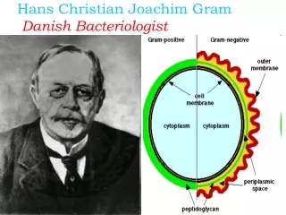
Dyes become Stains • With interest in the effects of dyes on living tissue. In 1884 the DanishmicrobiologistHans Christian Gram discovered that crystal violet irreversibly stains certain bacteria but can be washed from others. The dye has been widely used ever since for the Gram stain technique, which identifies bacteria as gram-positive (the stain is retained) or gram-negative (the stain is washed).
Gram staining a Important Technique • A staining technique used to classify bacteria; bacteria are stained with gentian violet and then treated with Gram's solution; after being decolorized with alcohol and treated with safranine and washed in water, those that retain the gentian violet are Gram-positive and those that do not retain it are Gram-negative
Gram positive Bacteria • Gram positive bacteria have a thick cell wall of peptidoglycan and other polymers. Peptidoglycan consists of interleaving filaments made up of alternating acetylmuramic acid and actylglucosamine monomers. In Gram positive bacteria, there are "wall teichoic acids". As well, between the cell wall and cell membrane, there is a "membrane teichoic acid".
Gram Negative Bacteria • Gram negative bacteria have an outer membrane of phospholipids and bacterial Lipopolysaccharides outside of their thin peptidoglycan layer. The space between the outer membrane and the peptidoglycan layer is called the periplasmic space. The outer membrane protects Gram negative bacteria against penicillin and lysozymes.
Definition of Gram stain • A method of staining bacteria using a violet stain. The gram staining characteristics (denoted as positive or negative). A heat fixed bacterial smear is stained with crystal violet (methyl violet), treated with 3% iodine/potassium iodide solution, washed with alcohol and counterstained. The method differentiates bacteria into two main classes, gram-positive and gram-negative.
Prefer to pick up colonies with loop and smear on Clean glass slide
Making a Smear • First prepare your slide. You do this by placing bacteria on a slide in a drop of water, allowing them to dry and then heat fixing them. Heating the slide kills the bacteria and makes sure that the bacteria a stuck to the slide and wont wash away during the staining procedure
Choosing a Right Smear • Before choosing a field for microscopic examination, it is important to look at the smear macroscopically • Note that the smear is easily visible in ordinary light
Requirements for Gram staining technique • Glass slides (25 by 75 mm), frosted ends desirable • b. 0.85% Nacl, sterile • c. Pasteur pipettes and wood applicator sticks, sterile • d. Microbiological loops, inoculating needles • e. Supplies for disposal of biological waste, including “sharps” • f. Bunsen burner • g. Immersion oil
Making Multiple smears in same slide – conserve resources • Making multiple smears make the optimal use of the slide. • Reduces the economic costs and saves the technical time.
Correct preparation • Smear preparation: Proper smear preparation should produce a monolayer of organisms sufficiently dense for easy visualization but thin enough to reveal characteristic morphological characteristics. Use clean, new glass slides. NOTE: When using the same pipette or swab, always inoculate culture media first
Steps in Gram Staining Procedure- Follow the Clock • 1On a rack, flood with filtered crystal violet 10 sec 2 Wash briefly in water to remove excess crystal violet • 3. Flood with Gram’s iodine 10 sec • 4. Wash briefly in water, do not let the section dry out. • 5. Decolourise with acetone until the moving dye front has passed the lower edge of the section • 6. Wash immediately in tap water • 7. Note : If the section appears too blue repeat steps 6 and 7 • 8. Counterstain with safranin 15 seconds
Observe the Following Under Oil Immersion lens • i. Relative amounts of PMN’s, mononuclear cells, and RBC's • ii. Relative amounts of squamous epithelial cells, bacteria • consistent with normal micro flora, which may indicate an improperly collected specimen • iii. Location and arrangements of microorganisms • Note any special character sticks
Microscopy: The Instruments • In a compound microscope the image from the objective lens is magnified again by the ocular lens. • Total magnification =objective lens ocular lens
Microscopy: The Instruments • Resolution is the ability of the lenses to distinguish two points. • A microscope with a resolving power of 0.4 nm can distinguish between two points ≥ 0.4 nm. • Shorter wavelengths of light provide greater resolution.
Optimal use of Microscopy • Gram stained preparations have to be observed with bright-field optics. Phase-contrast microscopy does not allow the recognition of true colours. Gram-positive bacteria may be seen under phase-contrast as red cells. Using bright-field optics, Gram-positive cells are purple or blue and Gram-negative pink due to counter stain with safranin..
Use Bright field optics • With bright-field optics colours can be discriminated best if the condenser iris is opened as far as possible without discomfort to the eyes
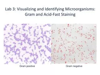
Colors makes the Difference in Gram staining • Bacteria that manage to keep the original purple dye have only got a cell wall - they are called Gram positive. • Bacteria that lose the original purple dye and can therefore take up the secondreddye have got both a cell wall and a cell membrane - they are called Gram negative.
Report as follows • If no microorganisms are seen in a smear of a clinical • specimen, report “No microorganisms seen.” • ii. If microorganisms are seen, report relative numbers and • Describe morphology. • Observe predominant shapes of microorganisms
Gram Stain Differentiates Gram positive from Gram negative • Differential Stains: Gram Stain
Nature of Morphology in guides early Diagnosis in uncommon diseases
Creating Library of Gram Stains • Drain or gently blot excess oil For slide libraries and teaching collections that will be stored for longer periods, immersion oil can be removed with xylene solution and the slides can be cover slipped using Per mount to prevent fading.
Value of Direct Smears • Guide the physician on initial choice of antibiotic, pending results of culture and sensitivity. • Judge specimen quality. • Contribute to selection of culture media, especially with mixed flora. • Provide internal quality control when direct smear results are compared to culture results.
Mixed Flora of Staphylococcus and Streptococcusdifferentiates morphology