Download
1 / 29
390 likes | 634 Vues
Structure of Viruses. Prepared By : Abdulrahman M. El-Sha'er Supervised By : DR. Abdelraouf Elmanama. SIZE & SHAPE. Viruses range from 20 to 300 nm in diameter; this corresponds roughly to a range of sizes from that of the

E N D
Structure of Viruses Prepared By : Abdulrahman M. El-Sha'er Supervised By : DR. Abdelraouf Elmanama
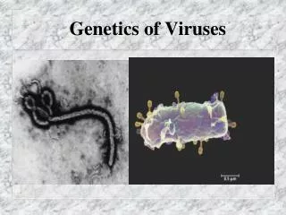
SIZE & SHAPE Viruses range from 20 to 300 nm in diameter; this corresponds roughly to a range of sizes from that of the largest protein to that of the smallest cell. Their shapes are frequently referred to in colloquial terms, eg, spheres, rods, bullets, or bricks, but in reality they are complex structures of precise geometric symmetry (see below). The shape of virus particles is determined by the arrangement of the repeating subunits that form the protein coat (capsid) of the virus.
VIRAL NUCLEIC ACIDS The viral nucleic acid (genome) is located internally and can be either single or double-stranded DNA or single- or double-stranded RNA. Only viruses have genetic material composed of single-stranded DNA or of single-stranded or double-stranded RNA. The nucleic acid can be either linear or circular.
The DNA is always a single molecule; the RNA can exist either as a single molecule or in several pieces. For example, both influenza virus and rotavirus have a segmented RNA genome. Almost all viruses contain only a single copy of their genome; ie, they are haploid. The exception is the retrovirus family, whose members have two copies of their RNA genome; ie, they are diploid.
VIRAL CAPSID & SYMMETRY The nucleic acid is surrounded by a protein coat called a capsid, made up of subunits called capsomers. Each capsomer, consisting of one or several proteins, can be seen in the electron microscope as a spherical particle, sometimes with a central hole. The structure composed of the nucleic acid genome and the capsid proteins is called the nucleocapsid. The arrangement of capsomers gives the virus structure its geometric symmetry.
Viral nucleocapsids have two forms of symmetry: (1) icosahedral, in which the capsomers are arranged in 20 triangles that form a symmetric figure (an icosahedron) with the approximate outline of a sphere; and (2) helical, in which the capsomers are arranged in a hollow coil that appears rodshaped. The helix can be either rigid or flexible. All human viruses that have a helical nucleocapsid are enclosed by an outer membrane called an envelope, ie, there are no naked helical viruses. Viruses that have an icosahedral nudeocapsid can be either enveloped or naked .
The advantage of building the virus particle from identical protein subunits is 2-fold: (1) it reduces the need for genetic information, and (2) it promotes self- assembly; ie, no enzyme or energy is required. In fact, functional virus particles have been assembled in the test tube by combining the purified nucleic acid with the purified proteins in the absence of cells, energy source, and enzymes.
VIRAL PROTEINS Viral proteins serve several important functions. 1- outer capsid proteins protect the genetic material and mediate the attachment of the virus to specific recep- tors on the host cell surface. 2- This interaction of the viral proteins with the cell receptor is the major determinant of species and organ specificity. Outer viral proteins are also important antigens that induce neutralizing antibody and activate cytotoxic T cells to kill virus-infected cells.
3- These outer viral proteins not only induce antibodies but are also the target of antibodies, ie, antibodies bind to these viral proteins and prevent ("neutralize") the virus from entering the cell and replicating. The outer proteins induce these immune responses following both the natural infection and immunization . Some of the internal viral proteins are structural (eg, the capsi d proteins of the enveloped viruses), whereas others are enzymes (eg, the polymerases that synthesize the viral mRNA).
If a virus has an envelope, then a matrix protein that mediates the interaction between the capsid proteins and the envelope proteins is present. Some viruses produce proteins that act as "superantigens" similar in their action to the superantigens produced by bacteria, such as the toxic shock syndrome toxin of Staphylococcus aureus .
Viruses known to produce superantigens include two members of the herpesvirus family, namely, Epstein-Barr virus and cytomegalovirus, and the retrovirus mouse mammary tumor virus.
VIRAL ENVELOPE In ad d ition to the capsid and internal proteins, there are two other types of proteins, both of which are associated with the envelope. The envelope is a lipoprotein membrane composed of lipid d erive d from the host cell membrane and protein that is virus-specific. Furthermore, there are frequently glycoproteins in the form of spike-like projections on the surface, which attach to host cell receptors during the entry of the virus into the cell. other protein, the matrix protein, mediates the interaction between the capsid proteins and the envelope.
The viral envelope is acquired as the virus exits from the cell in a process called “bu d d ing". The envelope of most viruses is derived from the cell's outer membrane, with the notable exception of her- pesviruses that derive their envelope from the cell's nuclear membrane. In general, the presence of an envelope confers instability on the virus. Enveloped viruses are more sensitive to heat, drying, detergents, and lipid solvents such as alcohol and ether than are nonenveloped (nucleocapsid) viruses, which are composed only of nucleic acid and capsid proteins.
An interesting clinical correlate of this observation is that virtually all viruses that are transmitted by the fecal-oral route (those that have to survive in the environment) do not have an envelope, that is, they are nake d nucleocapsid viruses. These include viruses suchas hepatitis A virus, poliovirus, coxsackievirus, echovirus, Norwalk virus, and rotavirus. In contrast, envelope d viruses are most often transmitted by d irect contact, such as by bloo d or by sexual transmission.
Examples of these include human immunodeficiency virus, herpes simplex virus type 2, and hepatitis B and C viruses. Other enveloped viruses are transmitted directly by insect bite, eg, yellow fever virus and West Nile virus, or by animal bite, eg, rabies virus. Many other enveloped viruses are transmitted from person to person in respiratory aerosol droplets, such as influenza virus, measles virus, rubella virus, respiratory syncytial virus, and varicella-zoster virus.
If the droplets do not infect directly, they can dry out in the environ- ment, and these enveloped viruses are rapidly inactivated. Note that rhinoviruses, which are transmitted by respiratory droplets, are naked nucleocapsid viruses and can survive in the environment for significant periods. They therefore can also be transmitted by hands that make contact with the virus on contaminated surfaces. The surface proteins of the virus, whether they are the capsid proteins or the envelope glycoproteins, are the principal antigens against which the host mounts its immune response to viruses.
They are also the determinants of type specificity (often called the serotype). For example, poliovirus types 1, 2, and 3 are distinguished by the antigenicity of their capsid proteins. It is important to know the number of serotypes of a virus, because vaccines should contain the prevalent serotypes. There is often little cross-protection between different serotypes. Viruses that have multiple serorypes, ie, have antigenic variants, have an enhanced ability to evade our host defenses because antibody against one serotype will not protect against another serotype.
ATYPICAL VIRUSLIKE AGENTS There are four exceptions to the typical virus as de- scribed above : (1) Defective viruses are composed of viral nucleic acid and proteins but cannot replicate witho,ut a "helper" virus, which provides the missing function. Defective viruses usually have a mutation or a deletion of part of their genetic material. During the growth of most human viruses, many more defective than infectious virus particles are produced.
The ratio of defective to infectious particles can be as high as 100:1. Because these defective particles can interfere with the growth of the infectious particles, it has been hypothesized that the defective viruses may aid in recovery from an infection by limiting the ability of the infectious particles to grow.
(2) Pseudovirions contain host cell DNA instead of viral DNA within the capsid. They are formed during infection with certain viruses when the host cell DNA is fragmented and pieces of it are incorporated within the capsid protein. Pseudovirions can infect cells, but they do not replicate.
(3) Viroids consist solely of a single molecule of circular RNA without a protein coat or envelope. There is extensive homology between bases in the viroid RNA, leading to large double-stranded regions. viroids replicate but the mechanism is unclear. They cause several plant diseases but are not implicated in any human disease.
(4) Prions are infectious particles that are composed solely of protiens; ie, they contain no detectable nucliec acid. They are implicated as the cause of certain “ slow “ diseases called transmissible spongiform encephalopthieswich include such diseases as Creutzfldt-Jakob disease in humans and scrapie in sheep. Because neither DNA nor RNA has been detected in prions, they are clearly different from viruses . Furthermore, electron microscopy reveals filament rather than virus particles. Prions are much more resistant to inactivation by ultraviolet light and heat than are viruses. They are remarkably resistant to formaldehyde and nucleases. However, they are inacti-vated by hypochlorite, NaOH, and autoclaving.
Hypochlorite is used to sterilize surgical instruments and other medical supplies that cannot be autoclaved. Prions are composed of a single glycoprotein with a molecular weight of 27,000-30,000. With scrapie pri- ons as the model, it was found that this protein is en- coded by a single cellular gene. This gene is found in equal numbers in the cells of both infected and unin- fected animals. Furthermore, the amount of prion pro- tein mRNA is the same in uninfected as in infected cells. In view of these findings, posttranslational modi- fications of the prion protein are hypothesized to be the important distinction between the protein found in in- fected and uninfected cells.
There is evidence that a change in the conformation from the normal alpha-helical form (known as PrP c, or prion protein cellular) to the abnormal beta-pleated sheet form (known as PrP sc, or prion protein scrapie) is the important modification. The abnormal form then recruits additional normal forms to change their configuration, and the number of abnormal pathogenic particles increases. Although prions are composed only of proteins, specific cellular RNAs enhance the conversion of the normal alpha-helical form to the pathologic beta-pleated sheet form.
The function of the normal prion protein is unclear. There is some evidence that it is one of the signal transduction proteins in neurons and that it is a copper-binding protein.
The prion protein in normal cells is protease-sensitive, whereas the prion protein in infected cells is protease- resistant, probably because of the change in conformation.
The observation that the prion protein is the product of a normal cellular gene may explain why no immune response is formed against this protein; ie, tolerance occurs. Similarly, there is no inflammatory response in infected brain tissue. A vacuolated (spongiform) appearance is found, without inflammatory cells. Prion proteins in infected brain tissue form rod-shaped particles that are morphologically and histochemically indistinguishable from amyloid, a substance found in the brain tissue of individuals with various central nervous system diseases (as well as diseases of other organs).