Download
1 / 56
590 likes | 842 Vues
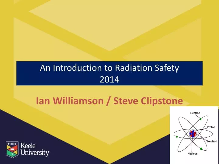
An Introduction to Radiation Safety 2014. Ian Williamson / Steve Clipstone. Alle Ding' sind Gift, und nichts ohn ' Gift; allein die Dosis macht , daß ein Ding kein Gift ist .

E N D
An Introduction to Radiation Safety2014 Ian Williamson / Steve Clipstone
Alle Ding' sind Gift, und nichtsohn' Gift; allein die Dosismacht, daßein Ding kein Gift ist. "All things are poison and nothing is without poison, only the dose permits something not to be poisonous." (Paracelsus, 1493 - 1541) Restricting Exposure
This course will cover • Main requirements of legislation • Types of radiation • Health effects • Management arrangements • Risk control measures
Aim The aim of the session is to • Introduce you to some basic radiation principles • Inform you of the arrangements and control measures in place to keep you safe when working with radiation • Not to make you an expert!
Early uses of radioactivity • Radium and thorium
Radiation Injuries • 1896 - first injuries due to radiation recorded • 1902 - first skin cancers seen • 1911 – 94 cases of skin carcinomas and sarcomas reported • H&S Legislation needed?
Legislation • Ionising Radiation Regulations 1999 (IRRs) General and specific duties & rules about safe working practices, control measures, assessments, roles and responsibilities; Health and Safety Executive (HSE) enforcement • Environmental Permitting Regulations 2010 Regulates the holding, storage, accumulation and disposal of radioactive material; Environment Agency (EA) Enforcement Replaced the RSA93 Act
The Ionising Radiation Regulations • Risk assessments • Control of exposure to ALARP • Maintenance of control measures • Dose limitation • Contingency Plans and Local Rules • RPA and RPS defined roles • Information. Instruction and Training • Co-operation between employers • Designation of areas
Dose Limits – For Workers • 1934 2 mSv per day or 730 mSv per year • 1937 2 mSv per day or 10 mSv per week • 1950 3 mSv per week or 150 mSv per year • 1956 1 mSv per week or 50 mSv per year • 1977 50 mSv per year • 2000 20 mSv per year
Annual Dose Limits – UK (IRRs) Women of reproductive capacity - exposure of abdomen limited to 13 mSv in any consecutive 3 month period. Women are legally obliged to inform their employer
Basic Radiological Safety Rules • All work must be risk assessed • You MUST work within the Local Rules and follow instructions • You MUST be a registered radiation worker • You MUST understand the instructions and comply with them – if in doubt ask
The ALARP Principle • Minimise the time you spend near a source • Maximise the distance between you and a source of radiation • Maximise the shielding between you and a source of radiation
Time • Before the work make sure you know and plan what you are going to do! Minimise the time • Practice the task beforehand • Do not linger in high dose rate areas
Distance • Avoid working or standing in high dose rate areas, whenever possible by moving away from the source of radiation • Use remote handling equipment • Observe from a separate area • Use minimal amounts / samples
Inverse Square Law DistanceRadiation Dose rate Double Reduced to ¼ Treble Reduced to 1/9 Quadruple Reduced to 1/16
Shielding • Use shielding provided where possible • Do not tamper with equipment or defeat interlocks • View behind protective screening • Make sure sealed sources are in good repair • PPE
Radiation Units Activity • Number disintegrations per second (Becquerel) – one Bq means one atom/nucleus decays and emits radiation every second • Characterised by the half life Absorbed dose • Mean energy per unit mass absorbed by any medium by any type of ionising radiation (Gray – Gy(or joules/kg)) Equivalent Dose • Dose allowing for type of radiation and effective biological damage (Sievert - Sv)- absorbed dose by weighting factor
Old/US Units • Rad 100 Rads = 1 Gray • Rem 100 Rem = 1 Sievert • Ci 1 Curie = 3.7 x 10¹⁰Bq (dps)
alpha • 2 protons + 2 neutrons tightly bound together - Helium nucleus • High energy but low penetrating power • Range in air only a few cm • Internal hazard
beta • Smaller than alpha • An electron (emitted from the nucleus) • Variable energy • Internal and external hazard
Gamma and x-rays • Electromagnetic radiation • Variable energy with shorter wavelengths • External hazard • Penetrating – range in air m to km • Gamma rays emitted from the nucleus • X-rays emitted from electron orbital shells
Radioactive Half-Life Not all of the atoms of a radioisotope decay at the same time, but they decay at a rate that is characteristic to the isotope. The rate of decay is a fixed rate called a half-life. The half-life of a radioisotope describes how long it takes for half of the atoms in a given mass to decay. Some isotopes decay very rapidly and, therefore, have a high specific activity. Others decay at a much slower rate– so decay at an “average rate” After two half-lives, there will be one quarter the original sample, after three half-lives one eighth the original sample, and so forth. It is an exponential decay process
= radioactive = stable, although not a precise figure After 1 half life half have decayed. There are 8 remaining 50% At start there are 16radioisotopes 100% After 2 half lives another half have decayed. There are 4 remaining 25% After 3 half lives another 2 have decayed. There are 2 remaining 12.5%
1 Half-Life 2 Half-Lives How can we work out the half-life of a radioisotope? We can plot a graph of activity against time
Routes of exposure Eye dose Inhalation Ingestion Skin dose Whole body dose Abdomen/Foetal Dose Extremity dose Injection
Routes of Entry • Ingestion • Inhalation • Puncture wounds or cuts • Absorption through the skin
Absorption of Nuclear Radiations The most massive of the radioactive emissions – alpha particles – have the shortest range. Due to their size they interact strongly with matter (lots of collisions with atoms) causing large amounts of ionization. This makes them very harmful to living tissue.
Absorption of Radiation Beta particles being smaller have a weaker interaction but can still cause ionization as they interact with the electrons surrounding atoms. Since gamma radiation is electromagnetic waves it is the most penetrating and least ionizing. However the deep penetration makes it dangerous to living tissue.
Biological Effects of Ionising Radiation • Health Effects are determined by the type and intensity of the radiation and the period of exposure.
Biological Effects • The occurrence of particular health effects from exposure to ionizing radiation is a complicated function of numerous factors including: • Type of radiation involved. All kinds of ionizing radiation can produce health effects. The main difference in the ability of alpha and beta particles and Gamma and X-rays to cause health effects is the amount of energy they have. Their energy determines how far they can penetrate into tissue and how much energy they are able to transmit directly or indirectly to tissues. • Size of dose received. The higher the dose of radiation received, the higher the likelihood of health effects. • Rate the dose is received. Tissue can receive larger dosages over a period of time. If the dosage occurs over a number of days or weeks, the results are often not as serious if a similar dose was received in a matter of minutes.
Part of the body exposed. Extremities such as the hands or feet are able to receive a greater amount of radiation with less resulting damage than blood forming organs housed in the torso. • The age of the individual. As a person ages, cell division slows and the body is less sensitive to the effects of ionizing radiation. Once cell division has slowed, the effects of radiation are somewhat less damaging than when cells were rapidly dividing. • Biological differences. Some individuals are more sensitive to the effects of radiation than others. Studies have not been able to conclusively determine the differences.
Radiation Effects • Direct ionisation • Structural cell damage, weakens links between atoms • Affects cellular function • DNA mutations • Indirect ionisation • Damage to chemical constituents, e.g. water • Formation of free radicals
Examples of various tissues and their relative radiosensitivities: High Radiosensitivity - Lymphoid organs, bone marrow, blood, testes, ovaries, intestines Fairly High Radiosensitivity- Skin and other organs with epithelial cell lining (cornea, oral cavity, esophagus, rectum, bladder, vagina, uterine cervix, ureters) Moderate Radiosensitivity - Optic lens, stomach, growing cartilage, fine vasculature, growing bone (note optic lens may move up to high radiosensitivity) Fairly Low Radiosensitivity - Mature cartilage or bones, salivary glands, respiratory organs, kidneys, liver, pancreas, thyroid, adrenal and pituitary glands Low Radiosensitivity - Muscle, brain, spinal cord Effects can take between 5 – 30 years
Radiation effects • Stochastic effects – somatic and hereditary effects • No safe dose or threshold – governed by chance • Deterministic effects – loss of function • There is no such thing as a safe level of radiation. A single electron could damage a cell irreversibly and initiate cancer However the likelihood of damage and the severity of damage increases with the amount of radiation.
Types of exposure • Acute exposure Takes place over a short period of time Usually high exposures • Chronic exposure Takes place over a long period of time Usually low level exposures
Stochastic effects Probability Effect, e.g. malignancy and hereditary effects Not immediately observable Dose probability increase as dose received increases
Deterministic Effects Threshold Severity Effect, e.g. cataracts, fetal damage, skin effects Large dose can be fatal Dose Degree of cells killed increases with dose impairing organ function
Deterministic Effects • 50 mSv body repairs itself • 1 Sv nausea and vomiting • 3 Sv Erythema, blistering and ulceration • 6 Sv LD50 depletion white blood cells, 50% population exposed die of infection death • 10 Sv severe depletion of cells lining intestine, death due to secondary infections
Radiation Detectors • Geiger counters, scintillation counters, ionisation chambers; • Count and sensitivity of the detector to interpret the readings
Monitors • Use portable radiation detectors to monitor laboratory or facility radiation levels • Use film badges or TLDs for retrospective personal dose monitoring • Calibrated contamination monitors are only valid for a particular type of radiation – there is no universal monitor
Work Areas • Controlled areas • Supervised Areas
Dosimetry • In controlled areas radiation dose is measured using dose meters or badges – you must wear them every time you enter a controlled area • You will be given specific instructions by your RPS
Restricting Exposure • All doses are kept to the ALARP principle Design – fail to safety and cannot be bypassed Engineered – shielded, fail to safety (interlocked), warning lights Administrative – Local Rules, supervision, disposal PPE – gloves , lab coats • Dose limits should not be exceeded
Risk Assessment All work requires a risk assessment where the risk is significant and foreseeable.IRRs require: Nature and source of ionising radiation to be used Estimated dose rates to anyone exposed Likelihood of contamination arising and being spread Results of previous monitoring if relevant Control measures and design features Requirement to designate areas and personnel Planned systems of work Estimated levels of airborne or surface contamination likely to be encountered • Requirement for PPE Possible accident situations, potential severity Consequences of failure of control measures Steps to limit consequences of accident situations
Local Rules • Brief and concise describing nature of work in the designated area • Identify key work instructions to restrict exposure • Covers normal circumstances and contingency plans • Contains realistic and achievable work instructions • Reviewed periodically to ensure effectiveness • Summary of arrangements for access restriction • Name / contact details of RPS should be in the local rules
Waste • Consult with your RPS regarding waste issues
Roles and responsibilities • Keele University – VC and the Committee Structure • Radiation Protection Advisor • University Radiation Protection Officer • Radiation Protection Supervisor • Registered Radiation Worker • Agencies / Regulatory bodies