Download
1 / 40
400 likes | 405 Vues
This chapter explains the concept of reflexes and the role of the peripheral nervous system in reflex activity. It covers different types of reflexes, the components of a reflex arc, and the importance of somatic reflex testing in assessing nervous system health.

E N D
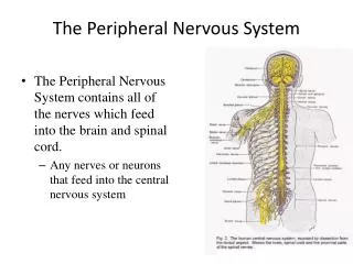
CHAPTER # 13(d) THE PERIPHERAL NERVOUS SYSTEM & REFLEX ACTIVITY
Reflexes • Inborn (intrinsic) reflex: a rapid, involuntary, predictable motor response to a stimulus • Learned (acquired) reflexes result from practice or repetition, • Example: driving skills
Reflex Arc • Components of a reflex arc (neural path) • Receptor—site of stimulus action • Sensory neuron—transmits afferent impulses to the CNS • Integration center—either monosynaptic or polysynaptic region within the CNS • Motor neuron—conducts efferent impulses from the integration center to an effector organ • Effector—muscle fiber or gland cell that responds to the efferent impulses by contracting or secreting
Stimulus Skin Interneuron 1 Receptor 2 Sensory neuron 3 Integration center 4 Motor neuron 5 Effector Spinal cord (in cross section) Figure 13.14
Spinal Reflexes • Spinal somatic reflexes • Integration center is in the spinal cord • Effectors are skeletal muscle • Testing of somatic reflexes is important clinically to assess the condition of the nervous system
Stretch and Golgi Tendon Reflexes • For skeletal muscle activity to be smoothly coordinated, proprioceptor input is necessary • Muscle spindles inform the nervous system of the length of the muscle • Golgi tendon organs inform the brain as to the amount of tension in the muscle and tendons
Muscle Spindles • Composed of 3–10 short intrafusal muscle fibers in a connective tissue capsule • Intrafusal fibers • Noncontractile in their central regions (lack myofilaments) • Wrapped with two types of afferent endings: primary sensory endings of type Ia fibers and secondary sensory endings of type II fibers
Muscle Spindles • Contractile end regions are innervated by gamma () efferent fibers that maintain spindle sensitivity • Note: extrafusal fibers (contractile muscle fibers) are innervated by alpha () efferent fibers
Secondary sensory endings (type II fiber) Efferent (motor) fiber to muscle spindle Efferent (motor) fiber to extrafusal muscle fibers Primary sensory endings (type Ia fiber) Extrafusal muscle fiber Muscle spindle Intrafusal muscle fibers Connective tissue capsule Sensory fiber Golgi tendon organ Tendon Figure 13.15
Muscle Spindles • Excited in two ways: • External stretch of muscle and muscle spindle • Internal stretch of muscle spindle: • Activating the motor neurons stimulates the ends to contract, thereby stretching the spindle • Stretch causes an increased rate of impulses in Ia fibers
Muscle spindle Intrafusal muscle fiber Primary sensory (la) nerve fiber Extrafusal muscle fiber Time Time (a) Unstretched muscle.Action potentials (APs) are generated at a constant rate in the associated sensory (la) fiber. (b) Stretched muscle.Stretching activates the muscle spindle, increasing the rate of APs. Figure 13.16a, b
Muscle Spindles • Contracting the muscle reduces tension on the muscle spindle • Sensitivity would be lost unless the muscle spindle is shortened by impulses in the motor neurons • – coactivation maintains the tension and sensitivity of the spindle during muscle contraction
Time Time (c) Only motor neurons activated. Only the extrafusal muscle fibers contract. The muscle spindle becomes slack and no APs are fired. It is unable to signal further length changes. (d) - Coactivation. Both extrafusal and intrafusal muscle fibers contract. Muscle spindle tension is main- tained and it can still signal changes in length. Figure 13.16c, d
Stretch Reflexes • Maintain muscle tone in large postural muscles • Cause muscle contraction in response to increased muscle length (stretch)
Stretch Reflexes • How a stretch reflex works: • Stretch activates the muscle spindle • IIa sensory neurons synapse directly with motor neurons in the spinal cord • motor neurons cause the stretched muscle to contract • All stretch reflexes are monosynaptic and ipsilateral
Stretch Reflexes • Reciprocal inhibition also occurs—IIa fibers synapse with interneurons that inhibit the motor neurons of antagonistic muscles • Example: In the patellar reflex, the stretched muscle (quadriceps) contracts and the antagonists (hamstrings) relax
Stretched muscle spindles initiate a stretch reflex,causing contraction of the stretched muscle andinhibition of its antagonist. The events by which muscle stretch is damped The sensory neurons synapse directly with alphamotor neurons (red), which excite extrafusal fibersof the stretched muscle. Afferent fibers alsosynapse with interneurons (green) that inhibit motorneurons (purple) controlling antagonistic muscles. 2 When muscle spindles are activatedby stretch, the associated sensoryneurons (blue) transmit afferent impulsesat higher frequency to the spinal cord. 1 Sensoryneuron Cell body ofsensory neuron Initial stimulus(muscle stretch) Spinal cord Muscle spindle Antagonist muscle 3a 3b Efferent impulses of alpha motor neuronscause the stretched muscle to contract,which resists or reverses the stretch. Efferent impulses of alpha motorneurons to antagonist muscles arereduced (reciprocal inhibition). Figure 13.17 (1 of 2)
Stretched muscle spindles initiate a stretch reflex,causing contraction of the stretched muscle andinhibition of its antagonist. The events by which muscle stretch is damped When muscle spindles are activatedby stretch, the associated sensoryneurons (blue) transmit afferent impulsesat higher frequency to the spinal cord. 1 Sensoryneuron Cell body ofsensory neuron Initial stimulus(muscle stretch) Spinal cord Muscle spindle Antagonist muscle Figure 13.17 (1 of 2), step1
Stretched muscle spindles initiate a stretch reflex,causing contraction of the stretched muscle andinhibition of its antagonist. The events by which muscle stretch is damped The sensory neurons synapse directly with alphamotor neurons (red), which excite extrafusal fibersof the stretched muscle. Afferent fibers alsosynapse with interneurons (green) that inhibit motorneurons (purple) controlling antagonistic muscles. 2 When muscle spindles are activatedby stretch, the associated sensoryneurons (blue) transmit afferent impulsesat higher frequency to the spinal cord. 1 Sensoryneuron Cell body ofsensory neuron Initial stimulus(muscle stretch) Spinal cord Muscle spindle Antagonist muscle Figure 13.17 (1 of 2), step 2
Stretched muscle spindles initiate a stretch reflex,causing contraction of the stretched muscle andinhibition of its antagonist. The events by which muscle stretch is damped The sensory neurons synapse directly with alphamotor neurons (red), which excite extrafusal fibersof the stretched muscle. Afferent fibers alsosynapse with interneurons (green) that inhibit motorneurons (purple) controlling antagonistic muscles. 2 When muscle spindles are activatedby stretch, the associated sensoryneurons (blue) transmit afferent impulsesat higher frequency to the spinal cord. 1 Sensoryneuron Cell body ofsensory neuron Initial stimulus(muscle stretch) Spinal cord Muscle spindle Antagonist muscle 3a Efferent impulses of alpha motor neuronscause the stretched muscle to contract,which resists or reverses the stretch. Figure 13.17 (1 of 2), step 3a
Stretched muscle spindles initiate a stretch reflex,causing contraction of the stretched muscle andinhibition of its antagonist. The events by which muscle stretch is damped The sensory neurons synapse directly with alphamotor neurons (red), which excite extrafusal fibersof the stretched muscle. Afferent fibers alsosynapse with interneurons (green) that inhibit motorneurons (purple) controlling antagonistic muscles. 2 When muscle spindles are activatedby stretch, the associated sensoryneurons (blue) transmit afferent impulsesat higher frequency to the spinal cord. 1 Sensoryneuron Cell body ofsensory neuron Initial stimulus(muscle stretch) Spinal cord Muscle spindle Antagonist muscle 3a 3b Efferent impulses of alpha motor neuronscause the stretched muscle to contract,which resists or reverses the stretch. Efferent impulses of alpha motorneurons to antagonist muscles arereduced (reciprocal inhibition). Figure 13.17 (1 of 2), step 3b
The patellar (knee-jerk) reflex—a specific example of a stretch reflex 2 Quadriceps(extensors) 3a 3b 3b 1 Patella Musclespindle Spinal cord(L2–L4) Tapping the patellar ligament excitesmuscle spindles in the quadriceps. 1 Hamstrings(flexors) Patellarligament 2 Afferent impulses (blue) travel to thespinal cord, where synapses occur withmotor neurons and interneurons. 3a The motor neurons (red) sendactivating impulses to the quadricepscausing it to contract, extending theknee. +– Excitatory synapseInhibitory synapse 3b The interneurons (green) makeinhibitory synapses with ventral horn neurons (purple) that prevent theantagonist muscles (hamstrings) fromresisting the contraction of thequadriceps. Figure 13.17 (2 of 2)
The patellar (knee-jerk) reflex—a specific example of a stretch reflex Quadriceps(extensors) 1 Patella Musclespindle Spinal cord(L2–L4) Tapping the patellar ligament excitesmuscle spindles in the quadriceps. 1 Hamstrings(flexors) Patellarligament +– Excitatory synapseInhibitory synapse Figure 13.17 (2 of 2), step 1
The patellar (knee-jerk) reflex—a specific example of a stretch reflex 2 Quadriceps(extensors) 1 Patella Musclespindle Spinal cord(L2–L4) Tapping the patellar ligament excitesmuscle spindles in the quadriceps. 1 Hamstrings(flexors) Patellarligament 2 Afferent impulses (blue) travel to thespinal cord, where synapses occur withmotor neurons and interneurons. +– Excitatory synapseInhibitory synapse Figure 13.17 (2 of 2), step 2
The patellar (knee-jerk) reflex—a specific example of a stretch reflex 2 Quadriceps(extensors) 3a 1 Patella Musclespindle Spinal cord(L2–L4) Tapping the patellar ligament excitesmuscle spindles in the quadriceps. 1 Hamstrings(flexors) Patellarligament 2 Afferent impulses (blue) travel to thespinal cord, where synapses occur withmotor neurons and interneurons. 3a The motor neurons (red) sendactivating impulses to the quadricepscausing it to contract, extending theknee. +– Excitatory synapseInhibitory synapse Figure 13.17 (2 of 2), step 3a
The patellar (knee-jerk) reflex—a specific example of a stretch reflex 2 Quadriceps(extensors) 3a 3b 3b 1 Patella Musclespindle Spinal cord(L2–L4) Tapping the patellar ligament excitesmuscle spindles in the quadriceps. 1 Hamstrings(flexors) Patellarligament 2 Afferent impulses (blue) travel to thespinal cord, where synapses occur withmotor neurons and interneurons. 3a The motor neurons (red) sendactivating impulses to the quadricepscausing it to contract, extending theknee. +– Excitatory synapseInhibitory synapse 3b The interneurons (green) makeinhibitory synapses with ventral horn neurons (purple) that prevent theantagonist muscles (hamstrings) fromresisting the contraction of thequadriceps. Figure 13.17 (2 of 2), step 3b
Golgi Tendon Reflexes • Polysynaptic reflexes • Help to prevent damage due to excessive stretch • Important for smooth onset and termination of muscle contraction
Golgi Tendon Reflexes • Produce muscle relaxation (lengthening) in response to tension • Contraction or passive stretch activates Golgi tendon organs • Afferent impulses are transmitted to spinal cord • Contracting muscle relaxes and the antagonist contracts (reciprocal activation) • Information transmitted simultaneously to the cerebellum is used to adjust muscle tension
2 1 Afferent fibers synapse with interneurons in the spinal cord. Quadriceps strongly contracts. Golgi tendon organs are activated. Interneurons Quadriceps (extensors) Spinal cord Golgi tendon organ Hamstrings (flexors) 3b 3a Efferent impulses to antagonist muscle cause it to contract. Efferent impulses to muscle with stretched tendon are damped. Muscle relaxes, reducing tension. + Excitatory synapse – Inhibitory synapse Figure 13.18
1 Quadriceps strongly contracts. Golgi tendon organs are activated. Interneurons Quadriceps (extensors) Spinal cord Golgi tendon organ Hamstrings (flexors) + Excitatory synapse – Inhibitory synapse Figure 13.18, step 1
2 1 Afferent fibers synapse with interneurons in the spinal cord. Quadriceps strongly contracts. Golgi tendon organs are activated. Interneurons Quadriceps (extensors) Spinal cord Golgi tendon organ Hamstrings (flexors) + Excitatory synapse – Inhibitory synapse Figure 13.18, step 2
2 1 Afferent fibers synapse with interneurons in the spinal cord. Quadriceps strongly contracts. Golgi tendon organs are activated. Interneurons Quadriceps (extensors) Spinal cord Golgi tendon organ Hamstrings (flexors) 3a Efferent impulses to muscle with stretched tendon are damped. Muscle relaxes, reducing tension. + Excitatory synapse – Inhibitory synapse Figure 13.18, step 3a
2 1 Afferent fibers synapse with interneurons in the spinal cord. Quadriceps strongly contracts. Golgi tendon organs are activated. Interneurons Quadriceps (extensors) Spinal cord Golgi tendon organ Hamstrings (flexors) 3b 3a Efferent impulses to antagonist muscle cause it to contract. Efferent impulses to muscle with stretched tendon are damped. Muscle relaxes, reducing tension. + Excitatory synapse – Inhibitory synapse Figure 13.18, step 3b
Flexor and Crossed-Extensor Reflexes • Flexor (withdrawal) reflex • Initiated by a painful stimulus • Causes automatic withdrawal of the threatened body part • Ipsilateral and polysynaptic
Flexor and Crossed-Extensor Reflexes • Crossed extensor reflex • Occurs with flexor reflexes in weight-bearing limbs to maintain balance • Consists of an ipsilateral flexor reflex and a contralateral extensor reflex • The stimulated side is withdrawn (flexed) • The contralateral side is extended
+ Excitatory synapse – Inhibitory synapse Interneurons Efferent fibers Afferent fiber Efferent fibers Extensor inhibited Flexor inhibited Arm movements Flexor stimulated Extensor stimulated Site of reciprocal activation:At the same time, the extensor muscles on the opposite side are activated. Site of stimulus: a noxious stimulus causes a flexor reflex on the same side, withdrawing that limb. Figure 13.19
Superficial Reflexes • Elicited by gentle cutaneous stimulation • Depend on upper motor pathways and cord-level reflex arcs
Superficial Reflexes • Plantar reflex • Stimulus: stroking lateral aspect of the sole of the foot • Response: downward flexion of the toes • Tests for function of corticospinal tracts
Superficial Reflexes • Babinski’s sign • Stimulus: as above • Response: dorsiflexion of hallux and fanning of toes • Present in infants due to incomplete myelination • In adults, indicates corticospinal or motor cortex damage
Superficial Reflexes • Abdominal reflexes • Cause contraction of abdominal muscles and movement of the umbilicus in response to stroking of the skin • Vary in intensity from one person to another • Absent when corticospinal tract lesions are present