Download
1 / 58
590 likes | 680 Vues
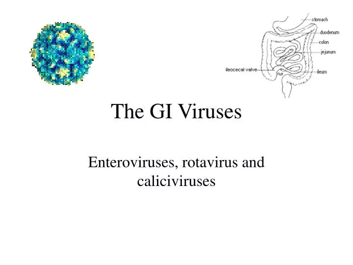
The GI Viruses. Enteroviruses, rotavirus and caliciviruses. Viruses that enter the body through the GI tract. Case number 1.

E N D
The GI Viruses Enteroviruses, rotavirus and caliciviruses
Case number 1 AK is a 22 year old bartender who presents to the ER in August with complaints of fever, chills, headache, and neck stiffness. He reports being well until a few days before admission when he noted the onset of fever, chills and general malaise. Over the past day his headache has become more prominent and his neck is now stiff. In the ER he is noted to have a temperature of 102oF, some pharyngeal erythema, and some mild neck stiffness. An LP is performed and shows a WBC count of 80 with 95% lymphocytes, normal protein and normal glucose. What’s going on?
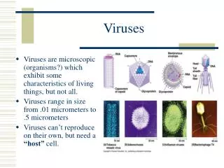
Molecular Biology • Belong to picornaviridae family • Aphthoviruses, enteroviruses, cardioviruses, rhinoviruses • 4 subgroups • Polioviruses, coxsackieviruses, echoviruses, newer enteroviruses • Non-enveloped, single-stranded, positive sense RNA viruses • Enter through the gastrointestinal tract
Epidemiology • World-wide in distribution • Non-polio enteroviruses have marked summer and fall seasonality in temperate zones • Spread person-person, by houseflies, wastewater and sewage • Fecal-oral spread facilitated by minimal clothing • Humans only significant natural reservoir • Physical exercise may impact manifestations of infection • Provocation polio • Myocarditis
Pathogenesis Minor viremia Mouth Gut- lymphatics Lymph node Virus v Heart Skin Lung Eye CNS Bloodstream Major viremia
Polioviruses • Cause of poliomyelitis • Humans only natural reservoir • Predilection for the central nervous system • Extensive necrosis of neurons in gray matter • Affects primarily motor and autonomic neurons • Anterior horn of spinal cord • Motor nuclei of pons and medulla
Epidemiology • In US prior to 1900 a usually subclinical disease of young infants • Milder disease than older children • Partial protection from maternal antibody • Improved hygiene- older kids affected • Increased paralytic disease • Epidemic 1950s • Vaccine 1955 • 2002- Polio eradicated in western hemisphere and Europe
Clinical Features • Range from inapparent illness to severe paralysis and death • 95% of infections are asymptomatic • Abortive poliomyelitis • Mild viral syndrome • Fever, headache, sore throat, listlessness • Normal neurologic exam • Lasts a few days • Nonparalytic poliomyelitis • Like abortive polio but signs of meningeal irritation • Full recovery
Spinal paralytic poliomyelitis • 0.1% of cases • Biphasic course • Minor illness- like abortive polio • Major illness- follows 2-5 days after recovery from minor illness • Abrupt illness- headache, fever, vomiting, neck stiffness, muscle pain for 1-2 days • Weakness and flaccid paralysis • Variable severity • Sensory loss rare
Bulbar Paralytic Poliomyelitis • Paralysis of muscles innervated by cranial nerves • Dysphagia, nasal speech, dyspnea • Cranial nerves 9 and 10 most commonly affected • Can involve vasomotor and respiratory centers • Rapid pulse • Hypoxia • Circulatory collapse
Polioencephalitis • Uncommon • Confusion and change in mental status • Most common in infants • Paralysis is spastic
Diagnosis • Viral isolation from throat- first week of illness • Isolation from stool for several weeks • Rarely isolated from CSF • Paired serology
Prevention • Two vaccine formulations • Oral Polio vaccine (OPV) • Live attenuated vaccine • Given orally • Excreted in feces- allows spread of vaccine to unimmunized individuals- herd immunity • Very rare- paralytic disease • Inactivated Polio vaccine (IPV) • Modified from original Salk vaccine • At least as immunogenic as OPV • Only vaccine used in US currently
Other Enteroviruses Coxsackieviruses, echoviruses, and newer enteroviruses
Clinical Manifestations • Do not usually cause symptomatic infections of the gastrointestinal system • Distributed worldwide • More prevalent in summer and autumn in temperate climates (June-October) • Most infections occur in children < 1 year
Central Nervous System • Aseptic meningitis • Prodrome- fever, chills, malaise, URI • Headache, fever, stiff neck, photophobia • 90% of viral aseptic meningitis in the community due to group B coxsackieviruses and echoviruses • CSF: 10-500 WBC, lymphocytes, nl to slightly elevated protein, nl glucose • PCR of spinal fluid usually reveals cause • Therapy is supportive
Encephalitis • Unusual manifestation of echovirus and coxsackievirus CNS infection • Accounts for 11-22% of viral encephalitis when you include polioviruses • Prognosis, except in infants, is excellent • Chronic meningoencephalitis • Seen in patients with acquired or congenital defects in B cell function • Echoviruses can be recovered from CSF for months-years • Try to prevent with monthly IG
Paralytic Infections • Occasionally associated with coxsackie and echovirus infections • Outbreaks of flaccid paralysis associated with coxsackievirus A7 and enterovirus 71 • Usually less severe than poliomyelitis • Paresis not permanent
Exanthems • Morbilliform rashes • Fine, erythematous, maculopapular rashes • Common in summer months • Rash appears simultaneously with fever and starts on face • Associated with echovirus 9
Roseoliform rashes • Discrete, nonpruritic, salmon-pink macules and papules on the face and upper chest • Prodrome of fever and pharyngitis • Rash appears after defervescence and lasts 1-5 days • Contagious especially amongst young children • Echovirus 16 most commonly associated
Hand, foot and mouth disease
Hand-foot-and-mouth disease • Distinctive vesicular eruption usually caused by coxsackie A16 or enterovirus 71 • Most common in children under age 10 • Fever and vesicles in the mouth and on the hands and feet • Can look like chickenpox but illness is generally milder
Generalized vesicular eruptions • Most frequently caused by coxsackievirus A9 and echovirus 11 • Lesions look like those of hand-foot-and-mouth but occur in crops on the head, trunk and extremities • Do not evolve into pustules or scabs (unlike chickenpox)
Herpangina • Vesicular rash involving pharynx and soft palate • Summer outbreaks of group A coxsackievirus • Fever, vomiting, myalgia and headache associated with prodrome
Respiratory Disease • Upper respiratory infections • Fever with sore throat, cough and coryza • Cause majority of summer colds in children • Coxsackieviruses A21 and A24; echovirus 11 • Epidemic pleurodynia • Acute disease with fever and sharp, spasmodic pain in chest/upper abdomen muscles • Fever peaks one after onset of pain spasm • Lasts 4-6 days usually but can persist for months
Myopericarditis • Inflammation of the myocardium and pericardium • Enteroviruses, especially group B coxsackieviruses, group A types 4 and 16 and echoviruses 9 and 22 account for 50% of all cases of acute myopericarditis • Virus appears to replicate in the myofibers leading to myofiber necrosis and focal inflammation
Special predilection for physically active adolescents and young adults • Males outnumber females 2:1 • Symptoms • URI in 70% followed by • Dyspnea • Chest pain- precordial, dull • Fever • Malaise • EKGs usually abnormal, cardiac enzymes elevated • Can lead to chronic congestive heart failure
Enterovirus infection of the newborn • Neonates are especially susceptible to severe enterovirus infection • Most serious infections appear to occur perinatally and probably are acquired from the mother • Lack of macrophage activity in the neonate is probably responsible for seriousness of infections
Clinical Manifestations • Biphasic illness • Mild non-specific symptoms between 3 and 7 days of life followed by 1-7 days of well-being • Generalized disease follows • Myocarditis with encephalitis- group B coxsackieviruses • Fulminant hepatitis- hypotension, bleeding, multiple organ failure- echovirus 11 • Diagnosis by PCR of urine, feces, blood, CSF • Treatment is supportive; pleconaril disappointing
Acute hemorrhagic conjunctivitis • Enterovirus 70 associated • Epidemic outbreaks of eye pain, swelling and subconjunctival hemorrhage • Highly contagious • Usually bilateral • Most cases resolve spontaneously
Case Number 1 • 22yo man with aseptic meningitis in August • Differential diagnosis • Enteroviruses • Other viruses • Rickettsiae • Lyme disease • Non-infectious- especially NSAIDs • PCR of spinal fluid positive for enterovirus
Case Number 2 • EM is an 8 month old girl who is brought into her pediatrician’s office in early March because of fever, vomiting, and diarrhea. Her mother reported that she had begun to have fever and vomiting 2 days previously and started having profuse watery diarrhea that day. She was enrolled in a daycare program and her mother reported that several other children in her class were sick with similar symptoms. On exam the baby was lethargic and had a temperature of 103oF, an elevated pulse, and normal blood pressure. Her mucous membranes appeared dry and her skin turgor was reduced.In passing Mom notes that Dad’s got a mild case of “the runs”.
Rotavirus • Microbiology • Reovirus family • Wheel-like appearance • Large, non-enveloped RNA viruses • Eleven segments of double stranded RNA • Reassortment occurs • Require RNA polymerase to make mRNA • Seven antigenic groups named A-G • groups A-C cause disease in humans • group A viruses account for most human disease worldwide.
Viral Proteins Yellow- outer capsid - vp7 Red spikes- hemagglutinin - vp4 Blue and green- inner capsid and core - vp1, vp2, vp3, vp6 Nonstructural proteins- NS53, NS34, NS35, NS28, NS26, NS12 - NS26 acts as enterotoxin
1. 2 3 5 7 4 6 8 9 Rotavirus replication 1. Viral entry via phagosome 2. Release from phagosome 3. Uncoating, release of RNA, and transcription into mRNA 4. Production of viral proteins 5. Viral RNA synthesis 6. Movement of viral proteins 7. Movement of core to ER 8. Assembly of viral particle 9. Release of viral particle
Pathogenesis • Spread by fecal-oral route • Virus enters and replicates in mature villus cells of the small intestine • Infection kills cells and loss of absorptive area ensues • Lactose intolerance common following infection • Enterotoxin may also contribute to diarrhea • Highly infectious and hardy • 1 pfu can cause disease • Not killed by many disinfectants
Epidemiology • Worldwide distribution • Most common cause of diarrhea requiring hospitalization in the world • Account for 10-20% of diarrhea-related deaths in children • Up to 120,000 hospitalizations in US/year • Seasonal in temperate climates • Occur in winter months in North America • Outbreaks start in the south west and move up to the north east by spring • Everyone infected by age 3
Estimated global distribution of the 800,000 annual deaths caused by rotavirus diarrhea
Clinical Features • Range from asymptomatic to severe diarrhea • First infection more severe than subsequent • Maximal disease incidence in infants 6-24 mos • Up to 30% of adult cases are symptomatic • Symptoms include • Fever • Nausea/vomiting • Watery diarrhea without blood/mucous • Dehydration/electrolyte imbalance lead to hospitalization and death
Diagnosis • Clinical- febrile infant with diarrhea in the winter • ELISA- detect rotavirus antigen in stool sample • PCR • Electron microscopy • Serology- epidemiological tool • Treatment • Replace fluids and electrolytes (oral or IV) • Early feeding- promote enterocyte regeneration • Do NOT give antidiarrheal agents
Prevention • Wash your hands • Chlorine containing disinfectants • Vaccine • Rotashield® • Live, oral vaccine • Rhesus-human recombinant • 15 cases of intussusception in first 10 months after licensure led to withdrawal