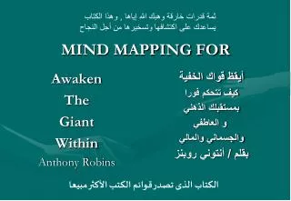
Download
1 / 6
60 likes | 65 Vues
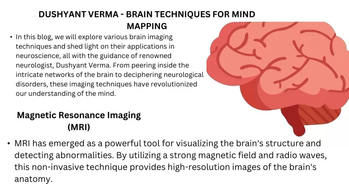
In this blog, we will explore various brain imaging techniques and shed light on their applications in neuroscience, all with the guidance of renowned neurologist, Dushyant Verma. From peering inside the intricate networks of the brain to deciphering neurological disorders, these imaging techniques have revolutionized our understanding of the mind.<br>

E N D
DUSHYANT VERMA - BRAIN TECHNIQUES FOR MIND MAPPING • In this blog, we will explore various brain imaging techniques and shed light on their applications in neuroscience, all with the guidance of renowned neurologist, Dushyant Verma. From peering inside the intricate networks of the brain to deciphering neurological disorders, these imaging techniques have revolutionized our understanding of the mind. Magnetic Resonance Imaging (MRI) • MRI has emerged as a powerful tool for visualizing the brain's structure and detecting abnormalities. By utilizing a strong magnetic field and radio waves, this non-invasive technique provides high-resolution images of the brain's anatomy.
Functional Magnetic Resonance Imaging (fMRI) • Dr.Dushyant Verma Shillong discusses how fMRI has revolutionized cognitive neuroscience, providing insights into language processing, memory, emotions, and various mental disorders such as schizophrenia and depression. Positron Emission Tomography (PET) • Dr. Dushyant Verma Shillong explains how PET scans help in understanding conditions like epilepsy, Alzheimer's disease, and brain tumors. PET is particularly valuable in studying neurotransmitter activity, revealing imbalances that contribute to mental illnesses and substance abuse disorders.
Electroencephalography (EEG) • Dr. Dushyant Verma Maharani Bagh highlights how EEG helps diagnose and monitor epilepsy, sleep disorders, and brain injuries. It also plays a crucial role in researching brain wave patterns associated with cognitive processes, allowing us to explore the intricacies of attention, learning, and brain-computer interfaces. Diffusion Tensor Imaging (DTI) • DTI is a specialized MRI technique that captures the brain's white matter tracts, enabling the visualization of neural connections. Dr. Dushyant Verma Maharani Bagh elucidates how DTI aids in mapping neural pathways and understanding conditions like multiple sclerosis, traumatic brain injuries, and neurodevelopmental disorders such as autism spectrum disorder.
Single-Photon Emission Computed Tomography (SPECT) • Dr. Dushyant Verma Southern Avenue explains how SPECT scans are particularly useful in diagnosing and monitoring conditions such as epilepsy, brain tumors, and psychiatric disorders. By detecting changes in blood flow, SPECT helps identify areas of the brain that may be affected by various neurological conditions. Magnetoencephalography (MEG) • This non-invasive technique allows researchers and clinicians to study brain function with high temporal resolution. Dr.Dushyant Verma Southern Avenue discusses how MEG is employed in mapping brain activity during cognitive tasks, localizing epileptic seizure sources, and understanding brain dynamics in various neurological disorders.
Optical Coherence Tomography (OCT) • Dr. Dushyant Verma highlights the importance of OCT in diagnosing and monitoring conditions such as multiple sclerosis, glaucoma, and optic neuropathies. By providing detailed images of the eye's layers, OCT assists in assessing the impact of neurological diseases on visual function. Molecular Imaging • Molecular imaging techniques, such as positron emission tomography (PET) with radiotracers, allow scientists to study specific molecules and receptors in the brain. Dr. Dushyant Verma Southern Avenue explains how molecular imaging helps investigate neurotransmitter systems, track disease progression, and evaluate the effectiveness of targeted treatments in neurology and psychiatry.
Future Directions • Dr. Dushyant Verma Maharani Bagh discusses the potential future advancements in brain imaging techniques. He explores emerging technologies like functional near-infrared spectroscopy (fNIRS) that offer portable and non-invasive brain imaging capabilities. Conclusion • Dr. Dushyant Verma Maharani Bagh has shared valuable insights into the applications of these techniques, emphasizing their crucial role in diagnosing neurological disorders, unraveling cognitive processes, and facilitating personalized treatments.As technology continues to evolve, we can look forward to further breakthroughs in mapping the mind, leading to enhanced understanding, improved patient care, and the potential for groundbreaking discoveries in neuroscience.