Download
1 / 25
321 likes | 637 Vues
Rh BLOOD GROUP SYSTEM. AHLS 311. HISTORY. Ab in serum of mother of stillborn child; responsible for the death of fetus? (1939, Levine and Stetson) Rb-derived Ab to Rhesus monkey RBCs reacts with 85% of human subjects; same Ab as reported by Levine? (1940, Landsteiner and Weiner)

E N D
Rh BLOOD GROUP SYSTEM AHLS 311
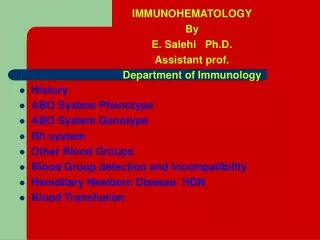
HISTORY • Ab in serum of mother of stillborn child; responsible for the death of fetus? (1939, Levine and Stetson) • Rb-derived Ab to Rhesus monkey RBCs reacts with 85% of human subjects; same Ab as reported by Levine? (1940, Landsteiner and Weiner) • Erythroblastosis fetalis (HDN) linked with Anti-Rh (1941, Levine et al)
NOMENCLATURE: 4 VERSIONS • Fisher Race • Suggested 3 sets of closely linked alleles (D and d, C and c, E and e) • Each gene (except d, which is an amorph) causes production of an Ag • Inherited from parents in linked fashion as haplotypes • See Tables 6-1 and 6-2
NOMENCLATURE • Weiner • Multiple alleles at 1 complex locus • 1 locus encodes for production of an agglutinogen which has 3 factors (antigens or epitopes) • Abs can recognize single or multiple factors • See Table 6-3
WEINER & FISHER-RACE TERMINOLOGY D = R 1 ( C) Z (both C & E ) 2 ( E ) 0 (neither C or E ) D c e D C E D C D c E d = r ‘( C) y (both C & E ) ‘’ ( E ) (neither C or E ) d c e d C E d C e d cE
NOMENCLATURE • Rosenfield • No genetic assumptions made • Numerical system • If listed alone, the Ag is present (Rh:1 = D Ag) • If listed with a “-”, the Ag is not present (Rh:1, -2, 3 = DcE) • If not listed, the Ag status was not determined • Adapts well to computer entry
NOMENCLATURE • Internatl. Soc. of Blood Transfusion • 6 digit number for each Ag specificity • First 3 indicate the blood group, eg., 004 = Rh • Last 3 indicates the Ag specificity, eg., 004001 = D Ag of Rh system • For recording of phenotypes, the system adopts the Rosenfield approach
Rh PHENOTYPING • Uses • Parentage testing • Predicting hemolytic disease of the newborn (HDN) • Confirmation of Rh Ab specificity • Locating compatible blood for recipients with Rh Abs • Protocol • Mix unknown RBCs with Rh antisera • Take tubes through phases (IS, heat/potentiator, AHG, CCC); record data • Use published frequencies and subject information to determine genotype
GENOTYPE FREQUENCIES • Dce (R1) 0.42 • dce (r) 0.37 • DcE (R2) 0.14 • Dce (R0) 0.04 • dCe (r’) 0.02 • dce (r”) 0.01 • DCE (Rz) <0.01 • dCE(ry) <0.01 The probability of 2 frequencies appearing together = the product of those 2 frequencies. For example, DCe/dce occurs with a frequency of 0.42 X 0.37 or 0.155.
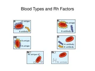
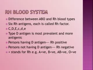
Rh ANTIGENS • Nonglycosylated proteins (A,B,H are CHOs) • Transmembrane molecules • D and CE are epitopes of proteins with 417 Aas that traverse the membrane 12 X • DNA sequences of D and CE differ by only 44 base pairs; CE, Ce, cd and cE are even more similar to D • Integral part of RBC membrane (Rhnull people have mild hemolytic anemia) • Density of Rh Ags on RBCs varies by phenotype (see Table 6-7)
D ANTIGEN VARIATIONS • Weak D • Some cells require addition of AHG (IDAT) to demonstrate agglutination with Anti-D • 3 mechanisms causing weak D expression • Genetic - inheritance of D genes which result in lowered densities of D Ags on RBC membranes • C trans - position effect; the D gene is in trans to the C gene, eg., Dce/dCe (C and D Ag arrangement causes steric hindrance weakening D expression) • D mosaic - 1 or more parts of the D Ag is missing; may result in production of Anti-D • People with weak D are considered Rh+ and receive Rh+ blood (except mosaics)
D ANTIGEN VARIATIONS • Enhanced D • When c and D are in double doses, eg., cDe/cDe, (C has limiting effect on expression of D) • D-- or D .. represent partial locus deletions; usually seen in consanguinous situations
D TESTING • Anti-D reagents • Saline-based - Low protein (fewer false positives); long incubation times; cannot convert to weak D testing • Protein-based - Faster, increased frequency of false positives; requires use of Rh control tube, converts to weak D testing • Chemically modified - “Relaxed” form of Anti-D in low protein medium; few false positives; saline control performed; converts to weak D testing • Blends of mAbs
D TESTING • Protocol • Add Anti-D to “D” tube; Rh control to “C” tube • Spin, read and record • If “D” is positive, cells are Rh positive • If “D” is negative, continue testing • Add 22% albumin and incubate for 20” at 37oC • Spin, read, and record • Wash 3 X in saline • Add AHG, spin, read, and record • If “D” is positive after heat/albumin or AHG cells are weak D positive; if negative, cells are Rh negative; “C” should always be negative • Add check cells to neg. tubes; spin, read & record
WEAK D Ag IN THE LAB • Differences from normal D expression • Quantitative (inherited weak D or position effects) • Qualitative (mosaic D; could produce Anti-D) • If cells are weak D, consider the person to be Rh + • Dwnot given to D negative recipients • D positives usually OK for Dw recipients • Dw mothers do not receive RhoGAM • Donors and expectant mothers should be tested for weak D; transfusion recipiencts +/- for weak D testing (Dw people may receive D negative blood)
OTHER ALLELES AND ANTIGENS • Weak C (Cw) • Not allelic to C and c (C and Cw usually seen together) • 2% of whites; very rare in blacks • Anti-Cw may be naturally occurring and shows dosage • f (ce) • When c & e are in cis, eg., dce/DCe • Combination Ag • Anti-f may be helpful in phenotyping
OTHER ALLELES AND ANTIGENS • Ce • When C and e in cis • Compound Ag • Ab helpful in phenotyping • G • Always found with C-positive RBCs; usually with D-positive cells • Anti G appears to bind to D, C, and G • Many others
ALLELIC DELETIONS • No Cc and/or Ee epitopes • DC-, Dc-, D-E, D-- • Enhanced or exalted D Ag expression • Rhnull (no Rh Ag expression at all) • ---/--- (double bar rr) • Or, because of independently inherited suppressor genes • If exposed to any Rh Ags, make Abs to those and to Rh 29 (“pan” or “total” Rh) • Causes a mild hemolytic anemia • Rhmod - weakened expression of all Rh Ags
Rh ANTIBODIES • Immune IgG Abs (IgG1 and IgG3 most important) • React optimally at 37oC or with AHG • Order of immunogenicity: D > c > E > C > e • Do not bind complement (RBC destruction by Rh Abs is extravascular)
Rh Abs: CLINICAL SIGNIFICANCE • Severe HDN • Severe transfusion reactions