Download
1 / 45
450 likes | 591 Vues
Chapter 13. Meiosis and Sexual Life Cycles. Overview: Variations on a Theme. living organisms are distinguished by their ability to reproduce their own kind Genetics = the scientific study of heredity and variation Heredity = the transmission of traits from one generation to the next

E N D
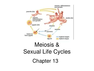
Chapter 13 Meiosis and Sexual Life Cycles
Overview: Variations on a Theme • living organisms are distinguished by their ability to reproduce their own kind • Genetics =the scientific study of heredity and variation • Heredity = the transmission of traits from one generation to the next • Variation = demonstrated by the differences in appearance that offspring show from parents and siblings
Inheritance of Genes • in a literal sense, children do not inherit particular physical traits from their parents • it is genes that are actually inherited • Genes are the units of heredity • made up of segments of DNA • passed to the next generation via reproductive cells called gametes (sperm and eggs) • somatic cells – any other cell other than the gamete or its precursor • each gene has a specific location called a locus on a certain chromosome
Comparison of Asexual and Sexual Reproduction • asexual reproduction - a single individual passes genes to its offspring without the fusion of gametes • produces an exact copy of the parent – a clone • done through fission or mitosis – following duplication of DNA • clone = a group of genetically identical individuals from the same parent • sexual reproduction - two parents give rise to offspring that have unique combinations of genes inherited from the two parents
APPLICATION Sets of Chromosomes in Human Cells • a life cycle = generation-to-generation sequence of stages in the reproductive history of an organism • a karyotype is an ordered display of the pairs of chromosomes from a cell • human somatic cells have 23 pairs of chromosomes • the two chromosomes in each pair are called homologous chromosomes, or homologs • chromosomes in a homologous pair have the same length, the same centromere position • also carry genes at the same loci controlling the same inherited characters TECHNIQUE 5 m Pair of homologousduplicated chromosomes Centromere Sisterchromatids Metaphasechromosome
Some Definitions You Know Already • sex chromosomes = X and Y • human females have a homologous pair of X chromosomes (XX) • human males have one X and one Y chromosome • remaining 22 pairs of chromosomes are called autosomes • each pair of homologous chromosomes includes one chromosome from each parent • a diploid cell (2n) has two sets of chromosomes • humans, the diploid number is 46 (2n = 46) • gamete (sperm or egg) contains a single set of chromosomes haploid (n) • humans, the haploid number is 23 (n = 23) • in an unfertilized egg (ovum), the sex chromosome is X • in a sperm cell, the sex chromosome may be either X or Y
Key Key Maternal set ofchromosomes (n 3) 2n 6 Paternal set ofchromosomes (n 3) • in a cell in which DNA synthesis has occurred - each chromosome has been replicated • each duplicated chromosome consists of two identical sister chromatids joined at a centromere Sister chromatidsof one duplicatedchromosome Centromere Two nonsisterchromatids ina homologous pair Pair of homologouschromosomes (one from each set)
Let’s Remind Ourselves Shall We? • chromosome = organized structure of DNA, protein and RNA found in the nucleus or nucleoid region • single piece of coiled DNA containing genes, regulatory elements and other nucleotide sequences • associated with DNA binding proteins – packaging of the DNA and control of gene expression • circular (prokaryotes) or linear (eukaryotes) • prokaryotic “chromosome” known as a genophore – not organized as chromatin • genes organized into operons – no introns • smaller, circular genophores = plasmids • circular “chromosomes” found in eukaryotic mitochondria and chloroplasts • true definition – DNA organized into chromatin • two forms in interphase – euchromatin (active form) and heterochromatin (inactive form) • replicated DNA condenses during the early stages of mitosis and meiosis to form two sister chromatids joined at a centromere 1. chromatid 2. centromere 3. p arm (short arm) 4. q arm (long arm)
centromere = point where the two sister chromatids join physical role – act as the site of assembly for the kinetochore kinetochore – complex group of proteins that attaches the centromere to the spindle microtubules and is responsible for chromatid separation (signals that all chromosomes are attached, aligned and are ready for separation) two kinetochore regions inner plate: associates with the centromere DNA sequences modified histone proteins that interact with DNA outer plate: associates with the microtubules Centromeres ** the sister chromatids are linked all along their length by cohesin proteins -arms are released during prophase until metaphase – only point of connection is the centromere
the heterochromatin of the centromere is key for interaction with the remaining cohesin protein complexes – keeps the chromatids together the tension receptors in the interzone sense the attachment of the kinetochore to microtubules cdc20 promotes the activation of the Anaphase Promoting Complex (APC) – a groups of proteins that initiates anaphase - triggers the separation of chromatids during anaphase the dynein motor proteins in the outer plate “walk” the separated chromatids to the opposite centrioles of the cell Centromeres – How’s this for cool?
cdc20 promotes the activation of the Anaphase Promoting Complex (APC) – a group of proteins that initiates anaphase - triggers the separation of chromatids during anaphase APC is a ubiquitin ligase – promotes the attachment of ubiquitin to protein targets degradation ubiquitin is attached to S and M cyclins – degradation results in transition from metaphase to anaphase ubiquitin is also attached to a protein complex made of proteins called securin & separase ubiquitin dependent degradation of securin “releases” separase from the complex separase now targets the cohesin protein complexes that link chromatids the dynein motor proteins in the outer plate “walk” the separated chromatids to the opposite centrioles of the cell APC ubiquination ubiquitin S and M cyclins Securin Separase Degradation of Securin Separase release Degradation Separase Metaphase Anaphase Transition Cohesin degradation Sister chromatid release It gets even cooler! Yes it does!
centromere = point where the two sister chromatids join two types: Point and Regional Point: smaller and more compact (e.g. yeasts) DNA sequence is necessary for the formation of the centromere bind to specific kinetochore proteins Regional: most centromeres (e.g. humans) DNA sequence contributes to formation of the centromere DNA of the centromere is heterochromatin not an actual defined sequence of DNA but is an array of repetitive sequences of “satellite DNA” that are similar to one another heterochromatin form of the satellite DNA is critical for the adherence of cohesin proteins in that region Centromeres 1. chromatid 2. centromere 3. p arm (short arm) 4. q arm (long arm)
centromere positions Metacentric: typical X shaped chromosome two arms (p and q) are equal in length humans – chr 1 & 3 Submetacentric: arms are unequal p is shorter Acrocentric: p arm is so short it is hard to observe humans: chr 13,14,15,21,22 and Y Telocentric: centromere is located at the end of the chromosome none in humans Holocentric: the entire length of the chromosome acts as the centromere plants and many invertebrates (nematodes) Centromeres chromosome 1 chromosome 13
the centromere position in humans is inherited daughter chromosomes will assemble their centromeres in the same positions as the parent chromosome epigenetic – nothing to do with DNA sequence role of H3 histone ? – H3 in the centromere region isreplaced with a related protein called Centromere Protein A/CENP-A (humans) essential for the assembly of kinetochore proteins at the centromere Centromeres
Haploid gametes (n 23) Key Behavior of Chromosome Sets in the Human Life Cycle Haploid (n) Egg (n) Diploid (2n) • Fertilization is the union of gametes - the sperm and the egg • the gametes have one set of chromosome - haploid • produced via meiosis from a diploid germ cell • gametes are the only types of human cells produced by meiosis rather than mitosis • has one set of chromosomes from each parent • the zygote produces somatic cells by mitosis and develops into an adult • fertilization and meiosis alternate in sexual life cycles to maintain chromosome number Sperm (n) MEIOSIS FERTILIZATION Ovary Testis Diploidzygote(2n 46) Mitosis anddevelopment Multicellular diploidadults (2n 46)
The Variety of Sexual Life Cycles Key Haploid (n) Haploid unicellular ormulticellular organism Haploid multi-cellular organism(gametophyte) • the alternation of meiosis and fertilization is common to all organisms that reproduce sexually • the three main types of sexual life cycles differ in the timing of meiosis and fertilization Diploid (2n) Gametes n n Mitosis Mitosis Mitosis Mitosis n n n n n n n n Spores n MEIOSIS FERTILIZATION n Gametes n Gametes MEIOSIS FERTILIZATION FERTILIZATION MEIOSIS Zygote 2n 2n 2n 2n Diploidmulticellularorganism(sporophyte) Zygote 2n Diploidmulticellularorganism Mitosis Mitosis Zygote (a) Animals (b) Plants and some algae (c) Most fungi and some protists
Key Animals Haploid (n) Diploid (2n) Gametes n n • gametes are the only haploid cells in animals • produced by meiosis • undergo no further cell division before fertilization • fuse to form a diploid zygote that divides by mitosis to develop into a multicellular organism n MEIOSIS FERTILIZATION Zygote 2n 2n Diploidmulticellularorganism Mitosis (a) Animals
Key Plants Haploid (n) Diploid (2n) Haploid multi-cellular organism(gametophyte) • plants and some algae exhibit an alternation of generations • their life cycle includes both diploid and haploid multicellular stages • diploid organism - called the sporophyte - makes haploid spores by meiosis • the spore grows (via mitosis) into a haploid organism called agametophyte • gametophyte bears the reproductive parts of the organism OR is the reproductive part • gametophyte makes haploid gametes by mitosis • fertilization of gametes produces a new diploid sporophyte Mitosis Mitosis n n n n n Spores Gametes MEIOSIS FERTILIZATION 2n 2n Diploidmulticellularorganism(sporophyte) Zygote Mitosis (b) Plants and some algae
Key Fungi Haploid (n) Diploid (2n) Haploid unicellular ormulticellular organism • in most fungi and some protists, the only diploid stage is the single-celled zygote • there is no multicellular diploid stage • the zygote produces haploid spores by meiosis • each haploid cell grows by mitosis into a haploid multicellular organism - fungus • the haploid adult produces gametes by mitosis Mitosis n Mitosis n n n n Gametes MEIOSIS FERTILIZATION 2n Zygote (c) Most fungi and some protists
depending on the type of life cycle, either haploid or diploid cells can divide by mitosis • in animals – diploid cells undergo mitosis so that the organism grows into a diploid organism • specialized diploid germ cells undergo meiosis to produce gametes • gamete fusion produces a new diploid organism • in plants & fungus – haploid spores undergo mitosis so the organism grows into a haploid organism (e.g. gametophyte) • haploid organism produces gametes via mitosis • gamete fusion produces a diploid organism that produces haploid spores via meiosis (e.g. sporophyte) • only diploid cells can undergo meiosis • in all three life cycles, the halving and doubling of chromosomes contributes to genetic variation in offspring
Meiosis reduces the number of chromosome sets from diploid to haploid • meiosis is preceded by the replication of chromosomes • just like mitosis • meiosis takes place in two sets of cell divisions, called meiosis I and meiosis II • the two half as many chromosomes as the parent cell • cell divisions result in four daughter cells, each with
Interphase Pair of homologouschromosomes indiploid parent cell Chromosomesduplicate Duplicated pairof homologouschromosomes Sisterchromatids Diploid cell withduplicatedchromosomes Meiosis I Homologouschromosomes separate Haploid cells withduplicated chromosomes Meiosis II Sister chromatidsseparate 2 1 Haploid cells with unduplicated chromosomes The Stages of Meiosis • after chromosomes duplicate, two divisions follow • Meiosis I (reductional division): homologs pair up and separate, resulting in two haploid daughter cells with replicated chromosomes • Meiosis II (equational division): sister chromatids separate • the result is four haploid daughter cells with unreplicated chromosomes
Telophase I andCytokinesis Anaphase I Metaphase I Prophase I Centrosome(with centriole pair) Sister chromatidsremain attached Chiasmata Sisterchromatids Centromere(with kinetochore) Spindle Metaphaseplate Cleavagefurrow Homologouschromosomesseparate Fragmentsof nuclearenvelope Homologouschromosomes Microtubuleattached tokinetochore Each pair of homologous chromosomes separates. Two haploid cells form; each chromosomestill consists of two sister chromatids. • Division in meiosis I occurs in four phases: • Prophase I • Metaphase I • Anaphase I • Telophase I and cytokinesis Chromosomes line upby homologous pairs. Duplicated homologouschromosomes (red and blue)pair and exchange segments;2n 6 in this example.
Prophase I • prophase I typically occupies more than 90% of the time required for meiosis • many similarities with mitosis prophase • chromosomes begin to condense within the nucleus • the centrioles migrate and the spindle begins to forms • BUT a unique event happens – synapsis = pairing of two homologous chromosomes • ends of the chromosomes attach to proteins in the nuclear envelope • other proteins in the nuclear matrix help the 2 chromosomes align gene by gene • the paired chromosomes are called a tetrad • synapsis is followed by crossing over http://highered.mcgraw-hill.com/sites/0072495855/student_view0/chapter28/animation__stages_of_meiosis.html
Outer Plate Microtubules Inner Plate MITOSIS Metaphase I • the nuclear envelope is gone and the spindle is completing its formation • tetrads line up at the metaphase plate - with one chromosome facing each pole • microtubules from one pole are attached to the kinetochore of one chromosome of each tetrad • microtubules from the other pole are attached to the kinetochore of the other chromosome • different from mitosis – spindle attached to the kinetochores of one chromosome
Anaphase I • pairs of homologous chromosomes separate • one chromosome moves toward each pole - guided by the spindle apparatus • sister chromatids remain attached at the centromere and move as one unit toward the pole
Telophase I and Cytokinesis • in the beginning of telophase I - each half of the cell has a haploid set of chromosomes • BUT each chromosome still consists of two sister chromatids • cytokinesis forms two haploid daughter cells • in animal cells - a cleavage furrow forms • in plant cells - a cell plate forms • no chromosome replication occurs between the end of meiosis I and the beginning of meiosis II because the chromosomes are already replicated
Telophase II andCytokinesis Metaphase II Prophase II Anaphase II During another round of cell division, the sister chromatids finally separate;four haploid daughter cells result, containing unduplicated chromosomes. • Division in meiosis II also occurs in four phases • Prophase II • Metaphase II • Anaphase II • Telophase II and cytokinesis • Meiosis II is very similar to mitosis Haploid daughtercells forming Sister chromatidsseparate
Prophase II • the spindle apparatus re-forms • in late prophase II, chromosomes move toward the metaphase plate • line up like mitosis Metaphase II • the sister chromatids are arranged at the metaphase plate • BUT - because of crossing over in meiosis I - two sister chromatids of each chromosome are no longer genetically identical • kinetochores of sister chromatids attach to microtubules extending from opposite poles • like mitosis
Anaphase II • the sister chromatids separate – like mitosis • the sister chromatids of each chromosome now move as two newly individual chromosomes toward opposite poles – like mitosis • sister chromatid cohesion allows sister chromatids of a single chromosome to stay together through meiosis I • protein complexes called cohesinsare responsible for this cohesion • in mitosis: cohesins are cleaved at the end of metaphase • in meiosis I (anaphase I): cohesins are cleaved along the chromosome arms in anaphase I separation of homologs and at the centromeres • in meiosis II (anaphase II) separation of sister chromatids
Anaphase II & Cohesins • sister chromatid cohesion allows sister chromatids of a single chromosome to stay together through meiosis I • protein complexes called cohesins are responsible for this cohesion • cohesins – complex of 4 protein subunits (SMC1, SMC3, SSC1 & SSC3) • 4 protein subunits form a ring-like structure that encircles both chromatids • forms during the S phase of interphase • functions: • 1. holds sister chromatids together during metaphase and regulates their separation during anaphase of mitosis or meiosis • 2. facilitates chromatid attachment to spindle – interacts with the kinetochore and helps position the chromosomes at the metaphase plate • 3. facilitates recombination • 4. other functions – e.g. transcriptional regulation
Anaphase II & Cohesins • cohesins are cleaved by the enzyme called separase • in mitosis: all cohesins are cleaved at the end of metaphase • in meiosis I (anaphase I): cohesins are cleaved along the chromosome arms in anaphase I & not at the centromere separation of homologs and at the centromeres • in meiosis II (anaphase II) separation of sister chromatids just like mitosis • mechanism: • in meiosis I – the cohesin complex is protected from separase when in tetrad formation • only one kinetochore of the two per chromosome is attached to a microtubule at metaphase I protection from separase & chromosome separation • in meiosis II & mitosis – both kinetochores in the chromosome contact a microtubule and are susceptible to separase action
Telophase II and Cytokinesis • just like mitosis • the chromosomes arrive at opposite poles • nuclei form, and the chromosomes begin decondensing • Cytokinesis separates the cytoplasm • at the end of meiosis, there are four daughter cells - each with a haploid set of unreplicated chromosomes (half the number as the parent cell) • BUT - each daughter cell is genetically distinct from the others and from the parent cell
A Comparison of Mitosis and Meiosis SUMMARY Property Mitosis Meiosis • Mitosis conserves the number of chromosome sets, producing cells that are genetically identical to the parent cell • Meiosis reduces the number of chromosomes sets from two (diploid) to one (haploid), producing cells that differ genetically from each other and from the parent cell Occurs during interphase beforemitosis begins Occurs during interphase before meiosis I begins DNAreplication One, including prophase, metaphase,anaphase, and telophase Two, each including prophase, metaphase, anaphase,and telophase Number ofdivisions Occurs during prophase I along with crossing overbetween nonsister chromatids; resulting chiasmatahold pairs together due to sister chromatid cohesion Does not occur Synapsis ofhomologouschromosomes Four, each haploid (n), containing half as manychromosomes as the parent cell; genetically differentfrom the parent cell and from each other Two, each diploid (2n) and geneticallyidentical to the parent cell Number of daughter cellsand geneticcomposition Role in the animal body Enables multicellular adult to arise fromzygote; produces cells for growth, repair,and, in some species, asexual reproduction Produces gametes; reduces number of chromosomesby half and introduces genetic variability among the gametes
MITOSIS MEIOSIS Parent cell MEIOSIS I Chiasma Prophase Prophase I Chromosomeduplication Chromosomeduplication Duplicatedchromosome Homologouschromosome pair 2n 6 Metaphase I Metaphase Anaphase I Telophase I AnaphaseTelophase Haploidn 3 Daughter cells ofmeiosis I MEIOSIS II 2n 2n Daughter cellsof mitosis n n n n Daughter cells of meiosis II SUMMARY http://highered.mcgraw-hill.com/sites/0072495855/student_view0/chapter28/animation__how_meiosis_works.html
Meiosis: A summary • Three events are unique to meiosis, and all three occur in meiosis l • Synapsis and crossing over in prophase I: Homologous chromosomes physically connect and exchange genetic information • At the metaphase plate, there are paired homologous chromosomes (tetrads), instead of individual replicated chromosomes • At anaphase I, it is homologous chromosomes, instead of sister chromatids, that separate
Genetic variation produced in sexual life cycles contributes to evolution • Mutations (changes in an organism’s DNA) are the original source of genetic diversity • mutation is a change in the DNA sequence that lasts through rounds of mitosis or meiosis (i.e. perpetuates in the daughter cells) • mutations create different versions of genes called alleles • gametes are produced through meiosis – each with a unique complement of alleles • reshuffling of alleles during sexual reproduction produces genetic variation
Origins of Genetic Variation Among Offspring • the behavior of chromosomes during meiosis and fertilization is responsible for most of the variation that arises in each generation • three mechanisms contribute to genetic variation • Independent assortment of chromosomes • Crossing over • Random fertilization
Independent Assortment of Chromosomes Possibility 2 Possibility 1 Two equally probablearrangements ofchromosomes atmetaphase I • Homologous pairs of chromosomes orient randomly at metaphase I of meiosis • red chromosomes from “Dad”; blue chromosomes from “Mom” • when you undergo meiosis – multiple possibilities exist • see possibility 1 vs. 2 • in independent assortment - each pair of chromosomes sorts maternal and paternal homologs into daughter cells independently of the other pairs • so there is a 50% chance a daughter cell at the end of meiosis will get the maternal chromosome and a 50% chance it will get the paternal homolog
Possibility 2 Possibility 1 Two equally probablearrangements ofchromosomes atmetaphase I Metaphase II • the number of combinations possible when chromosomes assort independently into gametes is 2n, where n is the haploid number • for humans (n = 23), there are more than 8 million (223) possible combinations of chromosomes Daughtercells Combination 1 Combination 2 Combination 3 Combination 4
Prophase Iof meiosis Nonsister chromatidsheld togetherduring synapsis Crossing Over Pair of homologs Chiasma • crossing over produces recombinant chromosomes, which combine DNA inherited from each parent • begins very early in prophase I, as homologous chromosomes pair up gene by gene • homologous portions of two nonsister chromatids trade places • contributes to genetic variation by combining DNA from two parents into a single chromosome Centromere TEM Anaphase I Anaphase II Daughtercells Recombinant chromosomes
in crossing over the nonsister chromatids exchange DNA segments • also known as genetic recombination • produces recombinant chromosomes • the segments that are exchanged are similar in sequence and are on the same locations on either chromosome • the crossed region is called a chiasma – region where the two chromosomes are physically joined • each tetrad usually has more than one • requires an enzyme called a recombinase • major source of genetic variation http://www.tokyo-med.ac.jp/genet/anm/mimov.gif
crossing over can be unequal major source of gene duplication and deletions between chromosomes “equal” cross over site
Random Fertilization • random fertilization adds to genetic variation because any sperm can fuse with any ovum (unfertilized egg) • the fusion of two gametes (each with 8.4 million possible chromosome combinations from independent assortment) produces a zygote with any of about 70 trillion diploid combinations • crossing over adds even more variation • each zygote has a unique genetic identity
The Evolutionary Significance of Genetic Variation Within Populations • Natural selection results in the accumulation of genetic variations favored by the environment • Sexual reproduction contributes to the genetic variation in a population, which originates from mutations