Download
1 / 62
650 likes | 938 Vues
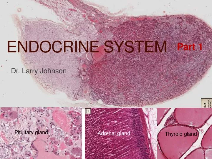
Endocrine system. Part 1. Dr. Larry Johnson. Pituitary gland. Adrenal gland. Thyroid gland. Objectives . Part 1 Distinguish between the neurohypophysis and the adenohypophysis and identify the cell types present in a slide or photomicrograph of the pituitary.

E N D
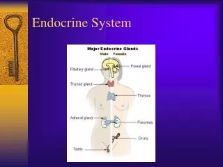
Endocrine system Part 1 Dr. Larry Johnson Pituitary gland Adrenal gland Thyroid gland
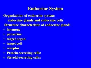
Objectives Part 1 • Distinguish between the neurohypophysis and the adenohypophysis and identify the cell types present in a slide or photomicrograph of the pituitary. • Identify thyroid follicles, follicular cells, colloid, capillaries and parafollicular cells. • Identify the capsule, chief cells, and oxyphil cells in the parathyroid gland. Part 2 • Identify the capsule, cortex, zona glomerulosa, zona fasciculata, zona reticularis, medulla, and chromaffin cells in the adrenal gland. • Identify the pinealocytes and corpora arenacea in the pineal gland. • Identify the islets of Langerhans in the pancreas
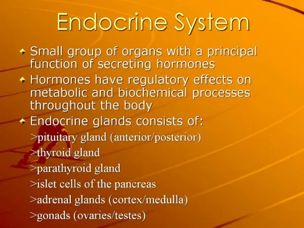
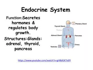
ENDOCRINE = INTERNAL SECRETION (without ducts) HORMONE = to AROUSE or SET in MOTION Endocrineglands are from endoderm
ORIGIN AND DISTRIBUTION OF EPITHELIUM ECTODERM - EPIDERMIS OF SKIN AND EPITHELIUM OF CORNEA TOGETHER COVERS THE ENTIRE SURFACE OF THE BODY; SEBACEOUS AND MAMMARY GLANDS ENDODERM - ALIMENTARY TRACT, LIVER, PANCREAS, GASTRIC GLANDS, INTESTINAL GLANDS ENDOCRINE GLANDS - LOSE CONNECTION WITH SURFACE MESODERM • ENDOTHELIUM - LINING OF BLOOD VESSELS • MESOTHELIUM - LINING SEROUS CAVITIES ECTODERM MESODERM ENDODERM
ORIGIN ENDODERM – ENDOCRINE GLANDS - LOSE CONNECTION WITH SURFACE ENDODERM
ADENOHYPOPHYSIS ORIGIN DIVISIONS I. PARS DISTALIS II. PARS TUBERALIS III. PARS INTERMEDIA RELATION TO HYPOTHALAMUS MICROSCOPIC ORGANIZATION I. CHROMOPHOBE CELLS II. CHROMOPHIL CELLS • ACIDOPHILS • BASOPHILS
Pituitary development http://php.med.unsw.edu.au/embryology/index.php?title=Endocrine_System_Development
We need to APPRECIATE THE DIVERSITY OF FUNCTIONS OF THE ENDOCRINE SYSTEM and to RECOGNIZE DIFFERENT ORGANS, UNIQUE FEATURES OF ORGANS, AND CELLS THAT MAKE THE ENDOCRINE SYSTEM
HORMONE = to AROUSE or SET in MOTION PHYSIOLOGICAL BLOOD LEVELS OF HORMONES compared to that for glucose GLUCOSE 10 -2 molar STEROID 10 -9 molar PEPTIDE 10 -12 molar GROWTH HORMONE (BLOOD LEVELS) 10 -13 molar = DWARF 10 -11 molar = GIANT
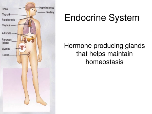
Pituitary gland (hypophysis) involvement in the neuroendocrine system Produces 9 hormones Reciprocal relations to other endocrine organs Neural and vascular connection to brain Location in key position for interplay between nervous and endocrine systems and establishment of neuroendocrine system
Pituitary gland PARS DISTALIS ADENOHYPOPHYSIS PARS TUBERALIS PARS INTERMEDIA PARS NERVOSA NEUROHYPOPHYSIS (PROCESSUS INFUNDIBULI) INFUNDIBULUM INFUNDIBULAR STEM MEDIAN EMINENCE OF THE TUBER CINEREUM
Pars distalis Pars intermedia Pars nervosa Pars intermedia Pars nervosa 79 Pars distalis Pars distalis Pars intermedia • 3 divisions of the pituitary gland: • Pars distalis • Pars intermedia • Pars nervosa
Slide 73: Pituitary (early carcinoma in posterior lobe) Infundibular stalk Carcinoma Pars intermedia Pars nervosa Pars distalis
Slide 73: Pituitary (early carcinoma in posterior lobe) Pars distalis Pars nervosa Acidophils Chromophobes Basophils Pituicyte nuclei
Herring bodies in pars nervosa of Hypophysis Pars nervosa
Slide 73: Pituitary (early carcinoma in posterior lobe) Pars intermedia with Rathke’s cysts Pars tuberalis
Slide 74: Pituitary (Masson’s trichrome) Infundibular stalk Carcinoma Pars nervosa Pars intermedia Pars distalis
Slide 73: Pituitary (early carcinoma in posterior lobe) Pars distalis Pars nervosa Chromophobes Acidophils Basophil Pituicyte nuclei Herring body
Slide 73: Pituitary (early carcinoma in posterior lobe) Pars intermedia with Rathke’s cysts Pars tuberalis
Thyroid gland • Follicle – no outlet • Colloid • Follicular cells • Capillaries in CT around follicle thyroid gland Colloid Capillary Follicular cells
Thyroid –parafollicular cells Parafollicular cells produce calcitonin
Slide 75: Thyroid gland Thyroid gland has many colloid-filled follicles. Thyroid hormones increase the number and size of mitochondria and stimulate mitochondrial protein synthesis, helping to enhance carbohydrate metabolism in cells. Parafollicular cells Secrete calcitonin – reduced blood calcium concentrations Follicular cells
Slide 39: Thyroid gland Parafollicular cells Thyroid gland with many colloid filled follicles Follicular cells
MEROCRINE SECRETION
Thyroid gland diseases Goiter - accumulation of thyroglobulin with iodine deficiency Graves disease – hyperthyroidism IgG immunoglobulin with long-acting thyroid stimulation
Endocrine secretions Stored in granules Stored extracellularly Immediate release with no storage pituitary thyroid adrenal Protein in cell Thyroglobulinoutside cell Steroid pass through cell in colloid of follicle
Slide 76: Parathyroid gland Chief (principal) cells vein Oxyphil cells Chief Oxyphils
Slide 76: Parathyroid gland Detached (artifact) connective tissue capsule Oxyphil cells Blood vessel Chief (principal) cells
Parathyroid – chief cells and oxyphils and rich vascular supply chief vein oxyphils vein chief
Parathyroid – chief cells Osteoporosis due to hyperparathyroidism chief
Endocrine system Part 2 Dr. Larry Johnson Pituitary gland Adrenal gland Thyroid gland
Objectives Part 1 • Distinguish between the neurohypophysis and the adenohypophysis and identify the cell types present in a slide or photomicrograph of the pituitary. • Identify thyroid follicles, follicular cells, colloid, capillaries and parafollicular cells. • Identify the capsule, chief cells, and oxyphil cells in the parathyroid gland. Part 2 • Identify the capsule, cortex, zona glomerulosa, zona fasciculata, zona reticularis, medulla, and chromaffin cells in the adrenal gland. • Identify the pinealocytes and corpora arenacea in the pineal gland. • Identify the islets of Langerhans in the pancreas
Releases of neurons associated with the adrenals (both direct and indirect)
BLOOD SUPPLY SINUSOIDS, MEDULLARY ARTERIES, ADRENAL VEIN • ZONA GLOMERULOSA • ZONA FASCICULATA • ZONA RETICULARIS
Adrenal Arterial and venous blood flow to the adrenal gland. • Peripheral arteries > cortical arteries > capillaries & sinusoids irrigating cortex > join medullary capillaries and arterioles > medullary fenestrated sinusoids with dual blood supply (arterial medullary blood and venous cortex blood) > medullary veins > suprarenal vein.
Human fetal adrenal cortical cellwith lots of SER and large spherical mitochondria with tubular cristae
Adrenal -cortex and medulla cortex medulla
Slide 77: Adrenal gland Cortex Medulla Zona reticularis Zona fasciculata Zona glomerulosa Capsule The zonaglomerulosa layer is regulated by pituitary adrenocorticotrophin hormone (ACTH).
Slide 77: Adrenal gland Chromaffin cells of medulla Sinusoidal blood channels Trabeculae of cortex Lipid droplets are abundant in these steroid-secreting cells. Cholesterol precursors for steroid hormones are stored in lipid droplets. Also SER would be abundant in these cells to provide the enzymes for steroid production.
Adrenal function The zonareticularis has a rich vascularization with wide capillaries. Aldosterone stimulates Na+resorption in: distal tubule of kidney gastric mucosa salivary glands sweat glands Cortisol - anti-inflammatory effects stabilizes lysomsomal membranes causes atrophy of lymphoid tissues throughout body decreases # of circulating lymphocytes
Adrenal function: blood pressure Slow but sustained effect on blood pressure Quick effect on blood pressure is by vasoconstriction of angiotensin II