Download
1 / 50
570 likes | 680 Vues
Acute Pericarditis Pericardial Effusion Tamponade. Outline. Anatomy of pericardium Overview of pericardial disease Etiology Clinical presentation Treatment. Anatomy. Normal amount of pericardial fluid: 15-50 cc Two layers:

E N D
Outline • Anatomy of pericardium • Overview of pericardial disease • Etiology • Clinical presentation • Treatment
Anatomy • Normal amount of pericardial fluid: 15-50 cc • Two layers: • Outer layer is the parietal pericardium and consists of layers of fibrous and serous tissue • Inner layer is visceral pericardium and consists of serous tissue only
Pericardium • Fibroelastic sac consisting of 2 layers • Visceral at epicardial side • Parietal at mediastinal side • Pericardial fluid formed from ultrafiltrate of plasma
Diseases of the Pericardium • Acute Fibrinous Pericarditis • Pericardial Effusion • Cardiac tamponade • Recurrent Pericarditis • Constrictive Pericarditis
Epidemiology of Acute Pericarditis • 0.1% of hospitalized patients • 5% of patients admitted to Emergency Department for non-acute myocardial infarction chest pain
Incidence • Exact incidence and prevalence are unknown • Diagnosed in 0.1% of hospitalized patients and 5% of patients admitted for non-acute MI chest pain • Observational study: 27.7 cases/100,000 population/year
Diagnostic Criteria • Chest pain: anterior chest, sudden onset, pleuritic; may decrease in intensity when leans forward, may radiate to one or both trapezius ridges • Pericardial friction rub: most specific, heard best at LSB • EKG changes: new widespread ST elevation or PR depression • Pericardial effusion: absence of does not exclude diagnosis of pericarditis • Supporting signs/symptoms: • Elevated ESR, CRP • Fever • leukocytosis
Diagnosis of Pericarditis: Presence of two of the following necessary 1) Chest pain • Sudden onset • localized to anterior chest wall • pleuritic • sharp • Positional: may improve if pt leans forward, worse with lying flat 2) Cardiac auscultation: Pericardial friction rub • Present in up to 85% of pts with pericarditis without effusion • friction of the two inflamed layers of pericardium, typically triphasic rub, heard with diaphragm of stethoscope at left sternal border 3) Characteristic ECG changes 4) Pericardial effusion
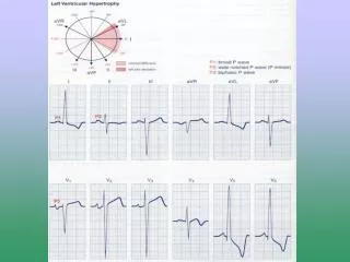
ECG Findings: 60% of patients • Stage 1: hours to days • Diffuse ST elevation -sensitive v5-v6, I, II • ST depression I/aVR • PR elevation aVR • PR depression diffuse -especially v5-v6 • PR change is marker of atrial injury • Stage 2: • Normalization
ECG changes over weeks • Stage 3: • Diffuse T wave inversions • ST segments isoelectric • Stage 4: • EKG may normalize • T wave inversions may persist indefinitely
STEMI or Pericarditis by ECG • ST elevation in pericarditis • Starts at J point • Rarely exceeds 5mm • Retains normal concavity • Non-localizing • Arrhythmias very unlikely in pericarditis (suggest myocarditis or MI)
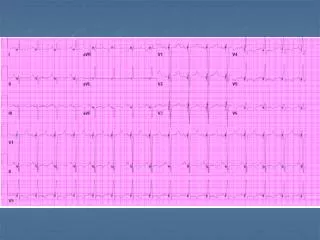
Acute Pericarditis • 51yo man with acute onset sharp substernal chest pain two days prior
EKG Electrocardiogram in acute pericarditis showing diffuse upsloping ST segment elevations seen best here in leads II, III, aVF, and V2 to V6. There is also subtle PR segment deviation (positive in aVR, negative in most other leads). ST segment elevation is due to a ventricular current of injury associated with epicardial inflammation; similarly, the PR segment changes are due to an atrial current of injury which, in pericarditis, typically displaces the PR segment upward in lead aVR and downward in most other leads.
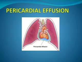
Pericardial Effusions *maximal width of pericardial stripe
Pericardial Effusion • Low voltage and Electric Alternans
Pericardial Effusion Cardiomegaly due to a massive pericardial effusion. At least 200 mL of pericardial fluid must accumulate before the cardiac silhouette enlarges.
Pericardial Effusion • M-Mode
Pericardial effusion • M-mode Cannot determine volume of accumulated fluid accurately
Treatment • Aspirin • NSAIDs • Colchicine: can reduce or eliminate need for glucocorticoids • Glucocorticoids: should be avoided unless required to treat patients who fail NSAID and colchicine therapy • Many believe that prednisone may perpetuate recurrences • Intrapericardialglucocorticoid therapy: sx improvement and prevention of recurrence in 90% of patients at 3 months and 84% at one year • Other immunosuppression • Azothoprine (75-100 mg/day) • Cyclophosphamide • Mycophenolate: anecdotal evidence only • Methotrexate: limited data • IVIG: limited data • Pericardiectomy: To avoid poor wound healing, recommended to be off prednisone for one year. Reserved for the following cases: • If >1 recurrence is accompanied by tamponade • If recurrence is principally manifested by persistent pain despite an intensive medical trial and evidence of serious glucocorticoid toxicity
Cardiac Tamponade • Normal amt of pericardial fluid = 20-50 mL • Tamponade occurs when lg or rapidly formed effusions inc’d pressure in the pericardial space throughout the cardiac cycle • During inspiration, RV volume inc’s & in tamponade, the RV is unable to expand into the maximally stretched pericardium L-ward bulging of the interventricular septum dec’d LVEDV dec’d cardiac output & dec’d SBP during inspiration
Tamponade • Pressure in pericardium exceeds s • Compressive effect in intrachamber • Diagnostic techniques • 2D looking for RA/RV collapse during diastole • M-mode for RA/RV collapse during diastole • Doppler of Mitral and Tricuspid inflow • Mitral inflow to decrease by 25% with inspiration • Tricuspid inflow increased by 40% with inspiration • IVC diameter fails to increase with inspiration
Etiology of Cardiac Tamponade • HIV, bacterial (incl mycobacterial), viral, fungal • CA - Esp lung, breast, Hodgkin’s, mesothelioma • Radiation tx • Meds - Hydralazine, Procainamide, INH, Minoxidil • Post-MI (free wall ventricular rupture, Dressler’s syndrome) • Connective tissue dzs – SLE, RA, Dermatomyositis • Uremia • Trauma • Iatrogenic – (eg, from TLC / PA Cath / TV pacemaker insertion, coronary dissection & perforation, sternal bx, pericardiocentesis, GE jnx surgeries) • Other - Pneumopericardium (d/t mech ventilation or gastropericardial fistula), Pleural effusions • Idiopathic
Clinical Presentation • Sxs • Chest Pain, dyspnea, near-syncope • Generally more comfortable sitting forward • Sxs c/w the underlying cause of tamponade • Physical Exam • Beck’s Triad - Elev’d JVP, hypotension, dec’d heart sounds • JVP w/ preserved x descent and dampened or absent y descent • Generally w/ narrow pulse pressure • Tachycardia, other signs of HF (tachypnea, diaphoresis, cool extremities, cyanosis, etc) • Pulsus paradoxus • Dec’d or absent cardiac impulse • +/- Friction rub
Pulsus “Paradoxus” • Dec in SBP > 10-12 mmHg w/ inspiration • Can also occur in pts w/ COPD, pulm dz, PTX, severe asthma • Can have tamponade w/o pulsus paradoxus • In pts w/ pre-existing elev’s in diastolic pressures and/or volume (eg, LV dysfnx, AI and ASD)
Diagnosis • Tamponade is a Clinical Diagnosis • Other Detection Methods • EKG • CXR • TTE • R Heart Cath • CT, MRI
EKG Findings • Common Findings • Sinus tachycardia • Non-specific ST segment and T wave changes • Changes assoc’d w/ acute pericarditis (incl diffuse STE & PR depression) • Other Findings • Dec’d voltage (non-specific and can also be d/t emphysema, infiltrative myocardial dz, PTX, etc) • Electrical alternans (specific but relatively insensitive for lg effusions) • 2/2 anterior-posterior swinging of the heart w/ each beat • Best seen in leads V2 to V4 • Combined P wave and QRS complex alternation (specific for cardiac tamponade)
CXR Findings Sudden inc in size of cardiac silhouette w/o specific chamber enlargement Effacement of the normal cardiac borders Development of a “flask” or “H2O-bottle” shaped heart
Lateral CXR Findings • May have (+) fat pad sign • Separation of mediastinal / retrosternal fat and epicardial fat by > 2 mm
Chest X ray • Normal in patients with acute pericarditis unless pericardial effusion is present • Enlarged cardiac silhouette • Requires 200cc of fluid
Tx of Cardiac Tamponade • If mild, can sometimes tx w/ medical mgmt • Including 1 or more of the following: NSAIDs, Colchcine, and/or steroids, depending on the suspected cause. • Require very close monitoring, including w/ serial TTEs and/or RHC
Tx of Cardiac Tamponade • Most require urgent/emergent pericardiocentesis • Closed pericardiocentesis • Generally in cath lab but can be at bedside • Subxiphoid approach under echo guidance is most common - minimizes risk & can assess completeness of fluid removal • Can alternatively use Fluoroscopic guidance • Pigtail catheter often left in place • Open Pericardiocentesis in the OR • May be best for loculated effusions, effusions containing clots or fibrinous material, and/or effusions that are borderline in size • Allow for bx and creation of a pericardial window for recurrent effusions • Bedside pericardiocentesis if pt is in extremis
Emergency Bedside Pericardiocentesis 16- or 18-gauge needle inserted at angle of 30-45° to the skin, near the left xiphocostal angle, aiming toward the L shoulder