Download
1 / 1
10 likes | 148 Vues
CERVICAL SPECIFIC PROTOCOL AND RESULTS FOR 139 MENIERE’S PATIENTS Michael T. Burcon, B.Ph., D.C. www.BurconChiropractic.com. LOGO. OBJECTIVES. RESULTS.

E N D
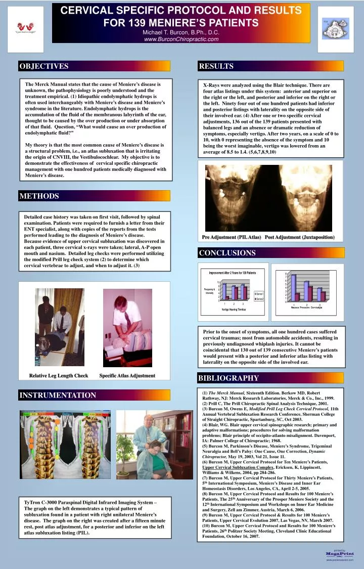
CERVICAL SPECIFIC PROTOCOL AND RESULTS FOR 139 MENIERE’S PATIENTS Michael T. Burcon, B.Ph., D.C. www.BurconChiropractic.com LOGO OBJECTIVES RESULTS The Merck Manual states that the cause of Meniere’s disease is unknown, the pathophysiology is poorly understood and the treatment empirical. (1) Idiopathic endolymphatic hydrops is often used interchangeably with Meniere’s disease and Meniere’s syndrome in the literature. Endolymphatic hydrops is the accumulation of the fluid of the membranous labyrinth of the ear, thought to be caused by the over production or under absorption of that fluid. Question, “What would cause an over production of endolymphatic fluid?” My theory is that the most common cause of Meniere’s disease is a structural problem, i.e., an atlas subluxation that is irritating the origin of CNVIII, the Vestibulocochlear. My objective is to demonstrate the effectiveness of cervical specific chiropractic management with one hundred patients medically diagnosed with Meniere’s disease. X-Rays were analyzed using the Blair technique. There are four atlas listings under this system: anterior and superior on the right or the left, and posterior and inferior on the right or the left. Ninety four out of one hundred patients had inferior and posterior listings with laterality on the opposite side of their involved ear. (4) After one or two specific cervical adjustments, 136 out of the 139 patients presented with balanced legs and an absence or dramatic reduction of symptoms, especially vertigo. After two years, on a scale of 0 to 10, with 0 representing the absence of the symptom and 10 being the worst imaginable, vertigo was lowered from an average of 8.5 to 1.4. (5,6,7,8,9,10) METHODS Detailed case history was taken on first visit, followed by spinal examination. Patients were required to furnish a letter from their ENT specialist, along with copies of the reports from the tests performed leading to the diagnosis of Meniere’s disease. Because evidence of upper cervical subluxation was discovered in each patient, three cervical x-rays were taken; lateral, A-P open mouth and nasium. Detailed leg checks were performed utilizing the modified Prill leg check system (2) to determine which cervical vertebrae to adjust, and when to adjust it. (3) Pre Adjustment (PIL Atlas) Post Adjustment (Juxtaposition) CONCLUSIONS Prior to the onset of symptoms, all one hundred cases suffered cervical traumas; most from automobile accidents, resulting in previously undiagnosed whiplash injuries. It cannot be coincidental that 130 out of 139 consecutive Meniere’s patients would present with a posterior and inferior atlas listing with laterality on the opposite side of the involved ear. Relative Leg Length Check Specific Atlas Adjustment BIBLIOGRAPHY (1) The Merck Manual, Sixteenth Edition. Berkow MD, Robert Rathway, NJ: Merck Research Laboratories, Merck & Co., Inc., 1999. (2) Prill C, The Prill Chiropractic Spinal Analysis Technique, 2001. (3) Burcon M, Owens E, Modified Prill Leg Check Cervical Protocol, 11th Annual Vertebral Subluxation Research Conference, Sherman College of Straight Chiropractic, Spartanburg, SC, Oct 2003. (4) Blair, WG. Blair upper cervical spinographic research; primary and adaptive malformations; procedures for solving malformation problems; Blair principle of occipito-atlanto misalignment. Davenport, IA: Palmer College of Chiropractic; 1968. (5) Burcon M, Parkinson’s Disease, Meniere’s Syndrome, Trigeminal Neuralgia and Bell’s Palsy: One Cause, One Correction, Dynamic Chiropractor, May 19, 2003, Vol 21, Issue 11. (6) Burcon M, Upper Cervical Protocol for Ten Meniere’s Patients, Upper Cervical Subluxation Complex, Ericksen, K, Lippincott, Williams & Wilkens, 2004, pp 284-286. (7) Burcon M, Upper Cervical Protocol for Thirty Meniere’s Patients, 5th International Symposium, Meniere’s Disease and Inner Ear Homeostasis Disorders, Los Angeles, CA, April 2-5, 2005. (8) Burcon M, Upper Cervical Protocol and Results for 100 Meniere’s Patients, The 25th Anniversary of the Prosper Meniere Society and the 12th International Symposium and Workshops on Inner Ear Medicine and Surgery, Zell am Zimmer, Austria, March 6, 2006. (9) Burcon M, Upper Cervical Protocol & Results for 100 Meniere’s Patients, Upper Cervical Evolution 2007, Las Vegas, NV, March 2007. (10) Burcon M, Upper Cervical Protocol and Results for 100 Meniere’s Patients, 26th Politzer Society Meeting, Cleveland Clinic Educational Foundation, October 16, 2007. INSTRUMENTATION TyTron C-3000 Paraspinal Digital Infrared Imaging System – The graph on the left demonstrates a typical pattern of subluxation found in a patient with right unilateral Meniere’s disease. The graph on the right was created after a fifteen minute rest, post atlas adjustment, for a posterior and inferior on the left atlas subluxation listing (PIL).