Download
1 / 19

E N D
Intussusception MiglenaKircheva PGY 1
QUESTION 1: A 1 year old boy with no significant PMH is brought to the ER with 6 hours history of fussiness and crying. The mother also notes that he was refusing to eat and vomited 4 times for the last 4 hours. The mother denies diarrhea, fever or any other symptoms. She is concerned because her baby is more sleepy and lethargic now. On physical exam the baby has tenderness in the right upper quadrant. What is the most common late presentation of this condition?
A. Weakness and lethargy • B. Bloody stools: ‘‘currant-jelly stools • C. Sausage-shaped mass on abdominal exam • D. Abdominaldistension; tenderness and intestine prolapse
ANSWER • A. Weakness and lethargy • B. Bloody stools: ‘‘currant-jelly stools • C. Sausage-shaped mass on abdominal exam • D. Abdominal distension; tenderness and intestine prolapse
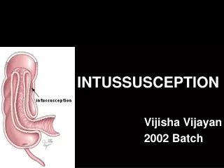
EXPLANATION A.) Weakness and lethargy In the classic description of intussusception, a child age 3 months to 3 years presents with the legs intermittently drawn up to the chest while crying, bloody stools, vomiting, and a sausage-shaped abdominal mass. Unfortunately, the classic presentation is not common. Many children present with lethargy or a change in mental status.
B) Bloody stools: ‘‘currant-jelly stools Blood is generally passed in the 1st 12 hr, but at times not for 1-2 days.60% of infants pass a stool containing red blood and mucus, the currant jelly stool. Some patients have only irritability and alternating or progressive lethargy. The classic triad of pain, a palpable sausage-shaped abdominal mass, and bloody or currant jelly stool is seen in <15% of patients with intussusception. The goal is to diagnose and treat intussusception prior to the evolution of ‘‘currant-jelly stools,’’ which is an indicator that significant bowel ischemia has occurred.
C) Sausage-shaped mass on abdominal exam Palpation of the abdomen usually reveals a slightly tender sausage-shaped mass, sometimes ill defined, which might increase in size and firmness during a paroxysm of pain and is most often in the right upper abdomen, with its long axis cephalocaudal. If it is felt in the epigastrium, the long axis is transverse. About 30% of patients do not have a palpable mass.
D) Abdominal distension; tenderness and intestine prolapse • Abdominal distention and tenderness develop as intestinal obstruction becomes more acute. On rare occasions, the advancing intestine prolapses through the anus.
QUESTION 2 What is the next BEST step in the management of this patient ? • A. Abdominal ultrasonography • B. Abdominal XR- plain film • C. Contrast enemas • D. Abdominal CT scan
ANSWER • A. Abdominal ultrasonography • B. Abdominal XR- plain film • C. Contrast enemas • D. Abdominal CT scan
EXPLANATION A) Abdominal ultrasonography US is the method of choice to detect intussusception. The sensitivity and specificity approaches 100% in the hands of an experienced technician. US can be used to monitor the success of a reduction procedure and does not expose the patient to radiation. A lack of perfusion in the intussusceptum detected with color duplex imaging may indicate the development of ischemia. Advantage of the US is that it can diagnose the rare ileo-ileal intussusception
B. Abdominal XR- plain film Abdominal XR is less sensitive and less specific than US for the diagnosis of intussusception. Early on, plain films are usually normal, but, as the disease progresses, up to 60% will show absence of air in the right upper and lower quadrants and evidence of soft tissue density. Radiographic features of intussusception include signs of intestinal obstruction, which may include massively distended loops of bowel with absence of colonic gas.
C. Contrast enemas Air, hydrostatic (saline), and, less often, water-soluble contrast enemas are the treatment of choice for a stable child, who was diagnosed with intussusception. Contrast enemas demonstrate a filling defect in the head of the contrast media where its advance is obstructed by the intussusceptum. A central linear column of contrast media may be visible in the compressed lumen of the intussusceptum, and a thin rim of contrast may be seen trapped around the invaginating intestine in the folds of mucosa within the intussusceptions (coiled-spring sign). Air reduction is associated with fewer complications and lower radiation exposure than traditional contrast hydrostatic techniques.
D. Abdominal CT scan An intussusception can be recognized on computed tomography , which may also identify the cause. However, CT can not be used to reduce the intussusception and can be time consuming in children who require sedation. Thus, CT is reserved for patients in whom the other imaging studies were not diagnostic
References Nelson Textbook of Pediatrics 19th Edition 2011; p 1287 : Intussusception Online resources: UpToDate: Intussusception in Children Pediatric emergency medicine secrets 2nd edition 2008