Download

1 / 95
980 likes | 1.48k Vues
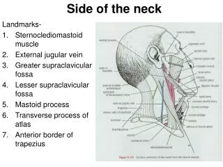
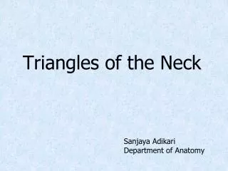
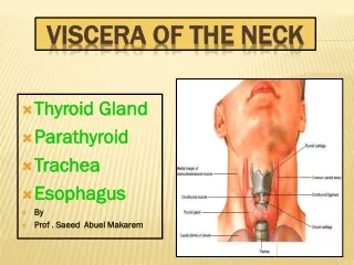
Dissection of the Neck. Surface Anatomy Surface landmarks : 1 suprasternal (jugular) notch(fossa), 2 hyoid bone, 3 cricoid cartilage, 4 thyroid cartilage, 5 sternocleidomastoid , 6 great supraclavicular fossa. Dissection of the Posterior Triangle of the Neck.

E N D
Surface Anatomy Surface landmarks :1suprasternal (jugular) notch(fossa), 2 hyoid bone, 3 cricoid cartilage, 4 thyroid cartilage, 5 sternocleidomastoid, 6 great supraclavicular fossa
Dissection of the Posterior Triangle of the Neck Boundaries and Contents of the Posterior Triangle. The boundaries of the posterior triangle are the sternocleidomastoid, the trapezius, and the clavicle. The triangle is roofed over by the investing layer of deep cervical fascia.
Make a midline skin incision from the point of the chin (symphysismenti) to the suprasternal notch. Continue the upper end of the incision backward and laterally along the lower border of the body of the mandible.
At the angle of the mandible, extend the incision backward, below the ear to the mastoid process and then along the superior nuchal line. From the lower end of the midline incision at the suprasternal notch, make an incision along the upper border of the clavicle to the acromion process.
Now carefully reflect the skin flaps laterally to expose the anterior and posterior triangles and the sternocleidomastoid muscle. Avoid damaging the underlying platysma muscle or the supraclavicular and accessory nerves.
If the arm has already been dissected, the skin will have been reflected from the back of the neck; the trapezius muscle will also been reflected laterally.
The superficial fascia is relatively thin and contains little fat; however, it possesses the platysma muscle. Carefully expose the paltysma muscle and note that its fibers arise from the superficial fascia covering the upper part of the pectoralis major muscle.
The fibers form a thin, broad sheet that passes upward and medially across the clavicle, covering the lower anterior part of the posterior triangle and the greater part of the anterior triangle. The muscle is inserted into the lower border of the body of the mandible and the angle of the mouth.
Divide the platysma along the upper border of the clavicle and reflect it upward and forward. Do not damage the underlying supraclavicular nerves and the external jugular vein that lie deep to it.
Locate the three supraclavicular nerves and note that they descend over the clavicle to supply the skin over the thoracic wall down as far as the sternal angle.
The lesser occipital nerve runs upward along the posterior border of the sternocleidomastoid muscle to be distributed to the skin over the auricle and mastoid process.
The great auricular nerve runs upward and forward to supply the skin over the angle of the mandible and the auricle.
Identify the external jugular vein and follow it superiorly to behind the angle of the mandible, where the external jugular is formed by the union of the posterior branch of the retromandibular vein and the posterior auricular vein. Trace the external jugular vein downward until it pierces the deep fascia. This vein varies considerably in size.
Now clean the sternocleidomastoid muscle and secure the cutaneous branches of the cervical plexus as they pierce the deep fascia at the posterior border of the sternocleidomastoid muscle.
The transverse cutaneous nerve crosses the sternocleidomastoid horizontally to supply the skin over the anterior triangle. The supraclavicular nerves, medial, intermediate, and lateral, descend over the clavicle, where they have already been secured.
Now continue to reflect the platysma as far as the mandible. At the angle of the mandible, examine the deep surface of the platysma and attempt to identify the cervical branch of the facial nerve, which emerges from the lower end of the parotid gland to supply the platysma muscle.
The boundaries of the posterior triangle are the sternocleidomastoid, the trapezius, and the clavicle. The triangle is roofed over by the investing layer of deep cervical fascia.
Examine the sternocleidomastoid and the trapezius muscles again. Identify and clean the inferior belly of the omohyoid muscle and note that this muscle subdivides the posterior triangle into an upper large occipital triangle and a lower smaller subclavian triangle (supraclavicular triangle).
Carefully clean the contents of the posterior triangle from the apex to the base. Secure the occipital artery as it crosses the apex. Identify the spinal part of the accessory nerve as it emerges from the posterior border of the sternocleidomastoid and follow it across the triangle on the levator scapulae until it disappears under the anterior border of the trapezius muscle.
Identify also the third and fourth cervical nerves, which run to the trapezius with the accessory nerve. Identify and clean the roots and trunks of the brachial plexus, which lie deeply in the lower anterior part of the posterior triangle.
The roots pass downward and laterally, entering the triangle between the scalenus anterior and medius muscles. The roots join one another to form the upper, middle, and lower trunks of the plexus, which continue laterally in front of the scalenus medius to enter the axilla in the interval between the clavicle and the first rib.
The anterior triangle is bounded by the lower margin of the mandible above, the sternocleidomastoid posterior, and the midline of the neck anteriorly.
The superficial fascia contains a variable amount of fat and the platysma. The platysma muscle should have been completely reflected upward to the lower margin of the mandible.
Identify again the cervical branch of the facial nerve as it leaves the parotid gland and runs forward, deep to the platysma, to supply it. Trace the transverse cutaneous nerve as it passes forward, across the sternocleidomastoidmuscle, to its distribution.
Identify and clean the anterior jugular vein as it lies in the superficial fascia near the midline and follow it inferiorly until it pierces the investing layer of deep cervical fascia. The anterior jugular vein is very variable in size.
Investing Layer of deep Cervical Fascia.Carefully remove the investing layer of the deep cervical fascia from the body of the hyoid bone above to the suprasternal notch below. Note that the fascia splits inferiorly into anterior and posterior layers, which are attached to the anterior and posterior border of the suprasternal notch.
Identify the suprasternal space that lies between the anterior and posterior layers of fascia and secure the jugular arch within the space that connects the anterior jugular veins to one another.
Now trace the anterior jugular vein laterally, deep to the sternocleidomastoid muscle, to where it joins the external jugular vein.
Subdivisions of the Anterior Triangle.The anterior and posterior bellies of the digastric muscle and the superior belly of the omohyoid muscle subdivide the anterior triangle into four subsidiary triangles, namely, the submental triangle, the digastric triangle, the carotid triangle, and the muscular triangle.
Carefully remove the remaining pierces of the investing layer of the deep cervical fascia that roofs over the anterior triangle of the neck.
Submental triangle. Identify the boundaries of this triangle. Note that it is bounded anteriorly by the midline of the neck, laterally by the anterior belly of the digastric muscle, and inferiorly by the body of the hyoid bone.
Clean the anterior belly of the digastric muscle and note that it arises from the body of the mandible. Note that the mylohyoid muscle forms the floor of the triangle. Identify within the triangle, if possible, the submental lymph nodes.
Digastric triangle.Confirm that the boundaries of the triangle are as follows: anteriorly, the anterior belly of the digastric muscle; posteriorly, the posterior belly of the digastric and the stylohyoid muscles; superiorly, the lower border of the body of the mandible.