Download
1 / 18
180 likes | 500 Vues
Hairs, Fibers, and Paint. Chapter 13. History of Hair Analysis. In 1883 Alfred Swaine Taylor and Thomas Stevenson wrote a forensic science text that included a chapter on hair. In 1910 Victor Balthazard and Marcelle Lambert published a comprehensive study of hair.

E N D
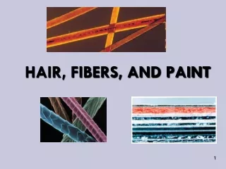
Hairs, Fibers, and Paint Chapter 13
History of Hair Analysis • In 1883 Alfred Swaine Taylor and Thomas Stevenson wrote a forensic science text that included a chapter on hair. • In 1910 Victor Balthazard and Marcelle Lambert published a comprehensive study of hair. • In 1934 Sydney Smith first used a comparison microscope to analyze hairs.
Morphology of Hair • Hair is part of the integumentary system. Figure 13-1 Cross section of skin showing hair growing out of a tubelike structure called the follicle.
Morphology of Hair • Hair grows from a hair follicle, extending from its root or bulb, and continuing into the shaft before terminating at the tip.
Morphology of Hair • The shaft of the hair is composed of three layers– the cuticle, cortex, and medulla.
imbricate spinous coronal Morphology of Hair • Cuticle- outside covering formed by overlapping scales - important feature for identifying human vs. animal hair and differentiating amongst different types of animal hairs e.g. imbricate- dog, spinous- rabbit, coronal- cat Figure 13-2 Scale patterns of various types of hair.
Morphology of Hair • Cortex- thickest layer made up of keratin, a type of protein; embedded with pigment granules whose color, shape and distribution provide important features for comparison
Morphology of Hair • Medulla- inner row of cells; humans may have a continuous, an interrupted, a fragmented or no medulla - The ratio of the diameter of the medulla relative to the diameter of the whole hair is called the medullary index (MI). - Human hair usually has an MI of 1/3 or less. - Animal hairs usually have an MI of ½ or more. Figure 13-4 Medulla patterns for various types of hair. Figure 13-5 Information on rabbit hair contained within the Forensic Animal Hair Atlas.
Morphology of Hair • Root- produces hair and allows it to grow • Hair growth occurs in three phases: anagen- growth phase that may last up to 6 years; root is attached to the follicle giving it a flame-shaped appearance; follicular tag allows for DNA analysis catagen- hair continues to grow, but at a decreasing rate for 2-3 weeks; root takes on an elongated appearance telogen- hair growth ends and hair is pushed out of the follicle over a 2-6 month period; root has a club-shaped appearance Figure 13-6 Hair roots in the anagen phase, categen phase, and telogen phase. - Hair grows approximately 1 cm per month.
Identification and Comparison of Hair • “Although animal hair can normally be distinguished from human hair with little difficulty, human hair comparisons must be undertaken with extreme caution and with an awareness of hair’s tendency to exhibit variable morphological characteristics, not only from one person to another but also within a single individual.”
Potential for Error • “Approximately 11 percent of the hairs (9 out of 80) in which FBI hair examiners found a positive microscopic match between questioned and standard/reference hairs were found to be non-matches when they were later subjected to DNA analysis.”
DNA Analysis • nuclear DNA- DNA present within the nucleus of a cell; inherited from both parents - obtained from follicular tags of hairs in the anagen or catagen phases • mitochondrial DNA- DNA present in organelles called mitochondria; only inherited from the mother - can be obtained from any hairs 1-2 cm in length
Collection and Preservation of Hair Evidence • Forensic hair comparisons usually involve either head hair or pubic hair. • The collection of 50 full-length hairs from all areas of the scalp will normally ensure a representative sampling of head hair. • A minimum collection of 24 full-length pubic hairs should cover the range of characteristics present in pubic hair. • Hair samples are also collected from the victims of suspicious deaths during an autopsy.
Types of Fibers • Natural fibers- derived from animal or plant sources e.g. animal- wool, cashmere, fur plant- cotton Figure 13-8 Photomicrograph of cotton fiber. • Manufactured fibers- derived from either natural or synthetic polymers, long chains of repeating subunits called monomers e.g. rayon (regenerated fiber), nylon (synthetic fibers) Table 13-1 Major Generic Fibers
Figure 13-10 Starch and cellulose are natural carbohydrate polymers consisting of a large number of repeating units or monomers. Figure 13-14 In the production of manufactured fibers, the bulk polymer is forced through small holes to form a filament in which all the polymers are aligned in the same direction.
Identification and Comparison of Manufactured Fibers • If the analyst can fit pieces of fiber together at their torn edges, then the evidence becomes individual. • Most often a comparison microscope is used to compare color, diameter and other features of fibers. Figure 13-11 Photomicrographs of synthetic fibers: cellulose triacetate and olefin fiber embedded with titanium dioxide particles. Figure 13-12 Cross-sectional shapes of fibers. Figure 13-13 A scanning electron photomicrograph of the cross section of a nylon fiber removed from a sheet used to transport the body of a murder victim. The fiber, associated with a carpet in Wayne Williams’s home, was manufactured in 1971 in relatively small quantities.
Identification and Comparison of Manufactured Fibers • The visible light microspectrophotometer is a convenient way for analysts to compare the colors of fibers through spectral patterns. • A more detailed analysis of the fiber’s dye composition can be obtained through a chromatographic separation. • Infrared spectrophotometryis a rapid and reliable method for identifying the generic class of fibers, as does the polarizing microscope. Figure 13-15 A photomicrograph of nylon fibers displaying interference colors when observed between the crossed polars of a polarizing microscope. Table 13-2 Refractive Indices of Common Textile Fibers
Collection and Preservation of Fiber Evidence • The investigator’s task of looking for strands of fibers often becomes one of identifying and preserving potential “carriers” of fiber evidence. • Relevant articles of clothing should be packaged carefully in separate paper bags. • If it is necessary to remove a fiber from an object, the investigator must use clean forceps, place it in a small sheet of paper, fold and label the paper, and place the paper packet inside another container.