Download
1 / 1

E N D
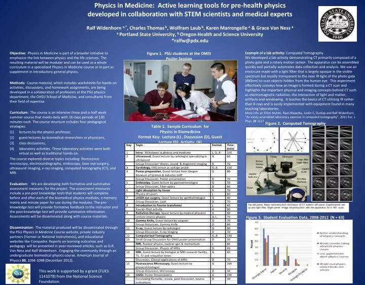
Physics in Medicine: Active learning tools for pre-health physics developed in collaboration with STEM scientists and medical expertsRalf Widenhorna,*, Charles Thomas b, Wolfram Laub b, Karen Marrongellea & Grace Van Ness aa Portland State University, b Oregon Health and Science University*ralfw@pdx.edu Example of a lab activity: Computed Tomography We developed a lab activity demonstrating CT primarily composed of a photo gate and a rotary motion sensor. The apparatus can be assembled quickly and partially automates data collection and analysis. We use an enclosure made with a light filter that is largely opaque in the visible spectrum but mostly transparent to the near IR light of the photo gate (880nm) to scan objects hidden from the human eye. This experiment effectively conveys how an image is formed during a CT scan and highlights the important physical and imaging concepts behind CT such as electromagnetic radiation, the interaction of light and matter, artifacts and windowing. It teaches the basics of CT utilizing IR rather than X-rays and is easily implemented with equipment found in many teaching laboratories. More info at: Elliot Mylott, Ryan Klepetka, Justin C. Dunlap and Ralf Widenhorn, “An easily assembled laboratory exercise in computed tomography”, 2011 Eur. J. Phys. 32 1227 Figure 1. PSU students at the OMSI Poster Session Objective: Physics in Medicine is part of a broader initiative to emphasize the link between physics and the life sciences. The resulting material will be modular and can be used as a whole curriculum in a specialized Physics in Medicine course or in part as supplement in introductory general physics. Methods: Course material, which includes worksheets for hands-on activities, discussions, and homework assignments, are being developed in a collaboration of professors at the PSU physics department, the OHSU School of Medicine, and consultants from their field of expertise. Curriculum: The course is an intensive three and-a-half week summer course that meets daily with 16 class periods of 130 minutes each. The course structure includes four pedagogical strategies: lectures by the physics professor, guest lectures by biomedical researchers or physicians, class discussions, laboratory activities. These laboratory activities were both virtual as well as traditional hands-on. The course explored diverse topics including: florescence microscopy, electrocardiography, endoscopy, laser eye surgery, ultrasound imaging, x-ray imaging, computed tomography (CT), and MRI. Evaluation: We are developing both formative and summative assessment measures for the project. The assessment measures include a pre-post knowledge tests that students will complete before and after each of the biomedical physics modules; a memory matrix and minute paper for use during the modules. The pre-knowledge test will provide formative feedback to the instructor and the post-knowledge test will provide summative information. Assessments will be disseminated along with course materials. Dissemination: The material produced will be disseminated through the PSU Physics in Medicine Course website, private industry partners (Vernier or National Instruments), and educational websites like Compadre. Reports on learning outcomes and pedagogy will be presented in peer-reviewed articles, such as G.R. Van Ness and Ralf Widenhorn. Engaging the community through an undergraduate biomedical physics course. American Journal of Physics 80, 1094-1098 (December 2012). Figure 2. Computed Tomography Table 1. Sample Curriculum for Physics in Biomedicine Format Key: Lecture (L) , Discussion (D), Guest Lecture (G), Activity, (A) Figure 3. Student Evaluation Data, 2008-2012 (N = 63) This work is supported by a grant (TUES 1141078) from the National Science Foundation.