Download
1 / 33
340 likes | 357 Vues
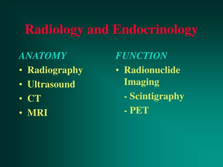
ANATOMY Radiography Ultrasound CT MRI. FUNCTION Radionuclide Imaging - Scintigraphy - PET. Radiology and Endocrinology. Radionuclide Imaging. Images metabolic pathways Pharmaceutical which mimics a component of a normal metabolic pathway is administered to the patient

E N D
ANATOMY Radiography Ultrasound CT MRI FUNCTION Radionuclide Imaging - Scintigraphy - PET Radiology and Endocrinology
Radionuclide Imaging • Images metabolic pathways • Pharmaceutical which mimics a component of a normal metabolic pathway is administered to the patient • Pharmaceutical radiolabelled so that its distribution in the patient can be visualised with a gamma camera
Ideal Radionuclide • emits gamma radiation at suitable energy for detection with a gamma camera (60 - 400 kev, ideal 150 kev) • should not emit alpha or beta radiation • half life similar to length of test • cheap • readily available
Ideal radiopharmaceutical • cheap and readily available • radionuclide easily incorporated without altering biological behaviour • radiopharmaceutical easy to prepare • localises only in organ of interest • t1/2 of elimination from body similar to duration of test
Thyroid - radiography • Little role • Thyroid mass diagnosed incidentally on chest radiograph • Thoracic inlet views may demonstrate tracheal compression
Thyroid - ultrasound • High resolution (5 - 10 MHz) • Confirms - mass is thyroid cystic or solid single or multiple • cannot distinguish solid carcinoma from solid dominant nodule • Not useful in hyperthyroidism
Thyroid - CT/MRI • Not as good as US at resolving lesions within the thyroid • Best tests for assessing mediastinal disease • CT better than MRI for calcification • MRI better than CT for distinguishing between fibrosis and residual tumour
Thyroid - scintigraphy 99m PERTECHNETATE Trapped but not organified Competes with iodide for uptake Cheap and readily available IODINE (123I or 131 I) Trapped and organified Better for retrosternal goitres Expensive, cyclotron generated RECENT (10 days) IODINE CONTRAST BLOCKS UPTAKE
Thyroid scintigraphy 99m Tc 123 NaI ADMIN iv po/iv PATIENT withdraw thyroid Rx PREP avoid high Iodine foods IMAGING 15 min pi 1-2hr pi 24 hr po
Hyperthyroidism RN uptake 1. Thyroid gland (>95%) Toxic nodular goitre Diffuse toxic goitre (Graves) Thyroiditis 2. Exogenous T3/4/iodine Iatrogenic Iodine - induced (XRay contrast, amiodarone)
Thyroid nodules Risk of malignancy Overall 10% US - cystic 0.3 - 10% US - solid ???? RNI - cold 16% RNI - hot 4% First line investigation: Cytology +/- US
RNI in thyroid disease • Investigation of hyperthyroidism • Location of ectopic thyroid tissue (congenital hypothyroidism, retrosternal goitre) • Little role in thyroid nodules
1ry Hyperparathyroidism Type % Adenomas Single 80 Hyperplasia Chief cell 15 Clear cell 1 Carcinoma 4
RN parathyroid imaging 99mTc / 201Tl 99mTc-MIBI subtraction scans early/late scans False positives: thyroid pathology False negatives: parathyroid hyperplasia Both good for ectopic parathyroids
Parathyroid imaging • US not good at finding ectopic glands • CT Contrast Surgical artifacts • MRI Good for localisation and ectopic glands
Imaging parathyroids Uncomplicated 1ry hyperparathyroidsim 90 -95% surgical success rate without imaging Recurrent/persistent hyperparathyroidism surgical success rate without imaging -50% with imaging - 90% (combined RNI + MRI)
Adrenal glands Cortex aldosterone cortisol adrenal androgens Medulla adrenalin
Adrenal glands • AXR - may show calcification • US - large masses only (unless neonatal) • CT - can detect small lesions - cannot distinguish metastases from non-functioning adenomas • MRI - small lesions - may distinguish mets from non-functioning adenomas
Adrenal cortical RNI • Radiolabelled cholesterol esters (75 Seleno-methylnorcholesterol, 131 I - 6B iodomethyl-19-norcholesterol) • Image at 4 and 7 days • > 50% difference in activity between sides is abnormal
RNI in Cushings syndrome ACTH-dependentCS bilat pituitary/ectopic ACTH -independent CS bilat nodular hyperplasia bilat adrenocortical adenoma uni Adrenocortical carcinoma bilat
Cushings syndrome Diagnosis - biochemistry Localisation - CT/MRI for 1. Pituitary ACTH-dependent 2. Ectopic ACTH-dependant 3. ACTH - independant RNI not usually necessary
RNI and Cushings syndrome Used for 1. Finding residual functioning adrenal remnants if recurrent disease after prior bilateral adrenalectomy 2. Somatostatin receptor scanning for ectopic ACTH from small bronchial carcinoid tumours
Primary aldosteronism • small tumours may not be seen with CT/MRI • RNI + dexamethasone suppression can find tumours < 1cm • Adrenal visualisation before 5 days is abnormal (bilateral/unilateral)
Adrenal medullary RNI Phaeochromocytoma Paraganglioma Neuroblastoma Ganglioneuroblastoma Ganglioneuroma
Adrenal medullary RNI • Metaiodobenzylguanidine (MIBG) -localises in catecholamine storage vesicles of adrenergic nerve endings - 123 I or 131 I • somatostatin receptor imaging 111 In octreotide
MIBG • phaeochromocytomas (95% sensitivity) • neuroblastoma (80 - 90% sens) • carcinoid • medullary thyroid carcinoma (MEN syndromes)
Phaeochromocytomas 10% malignant bilateral extra- adrenal paediatric
Phaeochromocytomas Diagnosis - biochemistry Localisation CT if > 2cm RNI to exclude - small tumours - bilateral adrenal - multifocal - metastases
‘Incidentalomas’ Incidental adrenal mass in patients undergoing abdominal imaging (2%) Q. Is it functioning? Is it benign or malignant?
Functioning ‘incidentalomas’ Diagnosis Clinical features Biochmistry Confirmation RNI
Non-functioning Non-functioning adenoma vs. metastasis • CT using attenuation values • MRI - chemical shift imaging
Radiology and Endocrinology Localisation not Diagnosis