Download
1 / 43
1.23k likes | 3.14k Vues
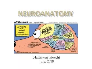
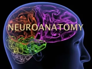
Neuroanatomy for Psychiatrists. Dr Rohit Shankar MBBS, MD, MRCPsych, CCT, PGC Cl. Research Consultant in Adult Developmental Neuropsychiatry. Why should we know any Neurology?. Brain Behaviour connection Man made divide 2000 years of togetherness

E N D
Neuroanatomy for Psychiatrists Dr Rohit Shankar MBBS, MD, MRCPsych, CCT, PGC Cl. Research Consultant in Adult Developmental Neuropsychiatry
Why should we know any Neurology? • Brain Behaviour connection • Man made divide • 2000 years of togetherness • Hippocrates (460-377BC) Humours theory and Triad of mental illness • Plato – divine inspired and physical inspired mental illness • Inter canon of the yellow emperor • Johann Christian Reil 1808 • Reintegration – biological underpinnings
Golden Rules • Adhere to the routine • A good History is more useful than a good examination • Usually well practiced testing would take 20 minutes then come back to any areas of deficits • Don’t ‘Scan’ before you ‘Can’ physically examine • Hoof beats are usually more likely to be from horses as opposed to Zebras, Hemiparesis is more likely from a stroke as opposed to an unwitnessed seizure
The Neurological Exam Motor System – Limb strength spasticity, flaccidity and fasciculation Abnormal movements – e.g.. Chorea and tremors Reflexes – DTRs – biceps, triceps, Quadriceps, Achilles Pathological reflexes – Babinski, frontal release signs Sensation – Position, vibration, stereognosis, Pain Cerebellar – Finger – Nose, Heel – Toe, Rapid alternating movements, Gait
The Neurological Exam Mental Status – GCS, orientation, Language, higher intellectual functions (arithmetic) Cranial Nerves – I Smell II Visual acuity, visual field, optic fundi Ocular motility nerves: III,IV,VI pupil size and reactivity, extra ocular motion cerebello-pontine angle nerves: V corneal reflex and facial sensation VII upper and lower facial muscle strength, taste VIII hearing Others: IX - XI articulation, palate movement, gag reflex XII tongue movements
DETAILS LIE IN BEHOLDER’S OBSERVATIONS! Detail of the Da Vinci's The Last Supper by Giacomo Raffaelli
Diagnostic Pathway Be Ritualistic The formulation: Symptoms, Signs, Localization and Diagnosis Localization: Where is the lesion? CNS, PNS or Muscles What is the lesion? Diffuse or Discrete Diagnosis: Common conditions arise commonly – Hoof beats are usually more likely to be from horses as opposed to Zebras Hemiparesis is more likely from a stroke as opposed to an unwitnessed seizure
Frontal Lobe Dysfunction • The primary motor cortex Contra lateral motor control • The medial frontal cortex Arousal and motivation – Abulic (Apathy & inattention) • The orbital frontal cortex Modulate Behaviour -Labile, euphoric, facetious, vulgar • The left postero-inferior frontal cortex (Broca's) Language – expressive Aphasia • The dorsolateral frontal cortex Working memory & dysexecutive syndrome
Parietal Lobe Dysfunction • The primary somatosensory cortex Integrates somesthetic stimuli for recognition and recall of form, texture, and weight - Contralateral astereognosis • Posterolateral - Postcentral gyrus visual-spatial relationships and proprioception • Midparietal lobe (dominant) calculation, writing, left-right orientation, and finger recognition - Gerstmann's syndrome • The nondominant parietal lobe Contralateral environmental awareness, drawing – Anosognosia, Hemiasomatognosia, spatial Apraxia
Temporal Lobe Dysfunction • Auditory perception, receptive components of language, visual memory, declarative (factual) memory, and emotion • Right temporal lobe lesions - interpret nonverbal auditory stimuli (e.g. music) • Left temporal lobe lesions interfere greatly with the recognition, memory, and formation of language • medial limbic - emotional parts & TLE
Occipital Lobe Dysfunction • Primary visual cortex and visual association areas • Anton Babinski Syndrome • Occipital Seizures – C/L Visual Hallucination • Prosopagnosia - Face blindness
Conscious pain, temperature, crude touch & pressure Lateral and an anterior tract Thalamus (all conscious sensations) projection to areas of the cerebral cortex
This tract carries unconscious proprioception (muscle sense) to the cerebellum which is responsible for muscle coordinationThey innervate the cerebellum on the same side
Corticospinal tract cerebral cortex – Localised voluntary motor controlTwo branches, the lateral and the anterior The lateral crosses in the medulla at the ‘pyramids’ The anterior does not crossCommon signs: DTR abnormalities, Motor Paresis, Babinski
The Basal Ganglia • Located Sub cortically • Modulates the Corticospinal tract • Regulates muscle tone, motor activity and generates postural reflex • Confined to the brain, no role on LMNs or Spinal Cord • Caudate Nucleus, Corpus Striatum, Lentiform Nucleus (Globus Pallidus + Putamen), Subthalamic Nuclei, Substantia Nigra
IC (white matter) runs between the CN and the LN = Corpus Striatum Artery of Stroke Pure damage to Basal Ganglia = No corticospinal symptoms, No neuropsychological dysfunction, No cognitive Dysfunction, contra lateral Result of biochemical not usually structural, B/L, slow progress Cerebrum + BG = inv Mov + cognitive &/or psychiatric Sx
Hippocampal Formation & Amygdala • Hippocampal Formation Dentate gyrus + the hippocampus proper + Subiculum Memory, spatial navigation and attention • Amygdala Via hypothalamus activates the ANS Activation of Neurotransmitters Emotional Learning – Conditioning Memory modulation Kluver Bucy Syndrome – Docility: diminished fear responses, dietary changes, Hyperorality, Hypersexuality, Visual Agnosia, Hypermetamorphosis: irresistible impulse to notice and react to everything, memory loss
Function of the Limbic System • Affective functions • Playful moods • Emotions and feelings, like wrath, fright, passion, love, hate, joy and sadness • self preservation
Dopamine Pathways HT VTA
Serotonin and Depression • Serotonin transmission - Caudal raphe nuclei and Rostal raphe nuclei is reduced in depression • Increasing the levels of serotonin in these pathways, by reducing serotonin reuptake = treatment
Serotonin in Schizophrenia • Dorsal raphe nuclei - Substantia Nigra • Rostral raphe nuclei - cerebral cortex, limbic regions and basal ganglia • The up-regulation of Serotonin pathways leads to the hypofunction dopamine pathways = negative symptoms • The serotonergic nuclei in the brainstem that give rise to descending serotonergic axons remain unaffected in schizophrenia
Brain Stem • Brain Stem: Midbrain, Pons, Medulla • Contains CNs, CS Tract and other ‘long’ Tracts • Positive evidence of localization and negative evidence of cerebral injury • Example – Diplopic but no effect on visual acuity or fields • Brain stem injures -Massive infarcts, Overdoses etc • Simultaneous damage of BS and Cerebrum RARE exceptions: MS, tumours etc
Cerebellum • Controls the coordination of movements/limbs – Ipsilateral • Muscle Hypotonia and Pendular DTRs • No obvious cognitive role • Intentional Tremor • Gait Ataxia, Scanning speech, tandem gait failure • Cognitive Impairment? • Alcohol – Thiamine, AIDS, toxins, Vitamin E, Phenytoin
Psychiatry and Neurology • Psychogenic Paresis and Hoover’s Sign • La Belle Indifference • MS • Sleep Disorders • Parkinsonism, Huntington, Wilson’s disease • Frontal Lobe issues, Dementia • Seizures of Non epileptic origin and NEADs, Sensory seizures
CASE STUDY 1 • An elderly man has left ptosis and a dilated and unreactive left pupil with external deviation of the left eye, right hemiparesis, right sided hyperactive DTRs and positive Babinski, no aphasia or hemianopia where is the lesion? • Cerebrum • Cerebellum • Pons • Midbrain • Medulla • None of the above
CASE STUDY 2 • A 20 year old woman reports having lost all vision in her right eye and right hemi-sensory loss. Pupil and DTRs are normal. She does not press down with her left leg while attempting to lift her right leg. where is the lesion? • Cerebrum • Cerebellum • Pons • Midbrain • Medulla • None of the above
CASE STUDY 3 • 50 yr old man with mild dementia has absent reflexes, loss of position and vibration sense and ataxia. Which areas are affected? • The CNS • The CNS and the PNS • The Cerebrum and the posterior columns • The ANS
CASE STUDY 4 • After having suffered from increasing severe depression for 3 years the psychiatrist finds the 55 year old woman to have right sided optic atrophy and left sided papilledema. Where is the lesion? • Occipital Lobe • Frontal Lobe • Parietal Lobe • Temporal Lobe • None of the above
QUESTION • Where is the primary damage in Wilson's disease, Huntington's Chorea and Choreiform Cerebral Palsy? • Extra pyramidal system • Pyramidal system • Entire CNS • Cerebellar outflow tracts • None of the above
SOME CORRECTIONS • EMI -2