Download
1 / 27
270 likes | 452 Vues
Lambda vectors and their replication Sonita Gafary Biochem 72020. Lambda was first discovered at the Pasteur Institute by Andre Lwoff when he observed strains of E. Coli. He showed that the cells of these bacterial strains carried bacteriophage in a dormant form (prophage).

E N D
Lambda vectors and their replication Sonita Gafary • Biochem 72020
Lambda was first discovered at the Pasteur Institute by Andre Lwoff when he observed strains of E. Coli. • He showed that the cells of these bacterial strains carried bacteriophage in a dormant form (prophage). • Phage can alternate between lysogenic (non-productive) and lytic (productive) growth cycles.
l Bacteriophage • Double stranded DNA molecule • 5' twelve-base-pair sticky ends (cos sites) • It is used as a cloning vector, accommodating fragments of DNA up to 15 kilobase pairs long. For larger pieces, the cosmid or YAC’s are used. • Will accept foreign DNA and still complete their life cycle. • Distinguish cells that have foreign and non foreign DNA. • Should replicate in host • Gene of interest can be identified and grown in large amounts. • Non essential genes can be removed and replaced by foreign gene.
Cont. • Should carry one or more selectable markers that identify the parent and recombinant vectors • Should have restriction sites in non-essential regions of DNA into which foreign DNA can be inserted • easy to make and maintain library
Enzymes needed: 1.Restriction enzymes: cuts the DNA at specific sequences to generate a set of fragments 2. DNA ligase: inserts DNA restriction fragments into replicating DNA molecules to produce recombinant DNA
1) Insertion vectors 2) Replacement vectors http://www.sh.lsuhsc.edu/gradcore/IDSP117/16 Lambda vectors cos cos EcoRI cos cos 20Kb EcoRI EcoRI
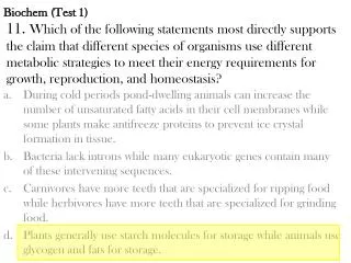
limitations • size of DNA to be introduced into the host cell • Problem: when making genomic libarary of large size (plants and mammals) only a portion of those fragments will be represented. If gene of interest is located in a large fragment, then you won’t be able to isolate that gene from the library. • Solution :use a vector that can accept large fragments of DNA
Vector types: 1. Plasmids- small circular DNA molecules which can replicate their DNA independently of their bacterial chromosome. They are found naturally in bacteria and replicate inside the bacterial cell. They can insert pieces up to 10kb(kilobases) or 100 to 10,000 base pairs. Examples: pBR322 and pUC18 2. Bacteriophage l-They are double stranded linear DNA vector. They replicate in E. Coli in the lytic or lysogenic mode. They can insert fragments up to 15kb. Examples are lgt10 and lZAP 3. Cosmids- are hybrid vectors of l phage and plasmids. They can replicate their DNA in the cell with a plasmid and be packaged like a phage. They can insert up to 50kb. 4. Yeast artificial chromosomes (YAC)- primarily used in genome sequencing projects. They host large inserts up to 1000kb.
What determines choice of vector? • Insert size • Vector size • Restriction sites • Cloning efficiency
-Central 1/3 is the “stuffer” fragment.-Segments of the lambda DNA are removed and a stuffer fragment is put in, this keeps the vector at a correct size
http://www.uic.edu/classes/phar/phar331/lecture6/ • Origin of replication is a DNA segment recognized by the cellular DNA-replication enzymes. Without replication origin, DNA cannot be replicated in the cell
http://www.uic.edu/classes/phar/phar331/lecture6 • Selective marker is required for maintenance of plasmid in the cell. Because of the presence of the selective marker the plasmid becomes useful for the cell. Under the selective conditions, only cells that contain plasmids with selectable marker can survive.
http://www.uic.edu/classes/phar/phar331/lecture6 • Many cloning vectors contain a multiple cloning site (DNA segment with several unique sites for restriction nucleases located next to each other)
http://www.uic.edu/classes/phar/phar331/lecture6 • Gene to be cloned can be introduced into the cloning vector at one of the restriction sites present in the cloning site.
Brock Biology of Microorganisms, 9th Edition (2000)Prentice Hall, Madigan, Martinko, Parker steps in cloning with l: • Isolate vector DNA and gene of interest • Cut both with restriction enzyme(EcoRI) • Connect two fragments of foreign DNA with DNA ligase. (recombinant DNA) • Package DNA by adding cell extracts containing head and tail proteins • Transfer recombinant molecules into host cell (transform) • Grow/select transformants: check recombinant phage for the presence of desired foreign DNA sequence by observing its genetic properties.
Molecular Biology of the Cell, 3rd Edition, Garland Publishing, Inc. 1983
-PL ( promoter) for transcription for the left side of l with N and cIII -PR (promoter) for right, including cro, cII and the genes encoding the structural proteins. -OL and OR is short non-coding region of genome, they control the promoters. -cI (repressor) protein of 236 a.a. which binds to OR and OL, preventing transcription of cro and N, but allowing transcription of OL, and the other genes in the left hand end. -cII and cIII encode activator proteins which bind to the genome. -Cro (66 aa) protein which binds to OR and OL, blocking binding of the repressor to this site to prevent lysogeny. -N codes an antiterminator protein and allows transcription from PL and PR. It also allows RNA polymerase to transcribe a number of phage genes, including those responsible for DNA recombination and integration of the prophage, as well as cII and cIII. -Q is an antiterminator similar to N, but only permits extended transcription from PR -Two Termination sites- One between N and CIII and other between cro and CII. http://www-micro.msb.le.ac.uk/224/Phages.html#Lambda
Life cycle of lambda • Virus enters cell • PL and PR gets activated • PL transcribes to make N protein • PR transcribes to make cro protein • Termination sites stop transcription but when enough N protein is made, transcription goes past these two stop sites (you can now make cIII and cII, replication proteins (O and P) and Q) • There are also termination sites next to Q protein. Q protein will allow transcription past this site. • If Cro protein blocks production of cI (goes lytic) • If cII and cIII activates transcription to make cI (goes lysogenic) • cI blocks PL and PR (stops transcription) by binding to OL and OR.
How do cells leave lysogeny cycle and go to lytic cycle? • By stress • ultraviolet irradiation of cells, this causes induction of a host cell protein, RecA whose normal function is to induce the expression of cellular genes which permit the cell to adapt to and survive in altered environmental conditions. RecA cleaves the cI repressor protein.
Which proteins determine the cycle? • Lysogenic cycle: cI proteins predominate • Lytic cycle: cro proteins predominate
DNA lambda replication • Initation of replication at the lambda origin requires “activation” by transcription starting from PR. • DNA replication is between O and P gene proteins. • Oril –Origin of phage l (with 4 binding sites adjacent to AT rich region)
O protein binds to lambda origin causing a structural change in the origin. P protein interacts with O protein Lambda proteins O and P form a complex with DnaB at the lambda origin (complex is inactive) This forms a spherical structure called an “O-Some” (~100bp of DNA) P protein (lambda’s) brings dnaB to the origin making the duplex larger (~160bp) The AT rich region becomes susceptible to nuclease attack (recognizes unpaired DNA), melting the DNA duplex. Shock proteins (dnaK, dnaJ and grpE gene) dissociate the oril O.P.dnaB complex to liberate dnaB dnaB initiates unwinding of duplex. Primase starts chain initations and polII starts elongation. DNA Replication, W.H. Freeman and Co. (1992) Kornberg,A.
-l circles multiply by theta form(q) and continues for 5-15 minutes after infection. -Rolling circles predominates after 15 minutes and produce linear concatemers (genomes linked end to end). -Packaging requires THF (termination host factor) provided by the host cell. DNA Replication, W.H. Freeman and Co. (1992) Kornberg,A.
References • http://www.uic.edu/classes/phar/phar331/lecture6/ • http://www.sh.lsuhsc.edu/gradcore/IDSP117/16 • http://www.gmu.edu/departments/biology/385-Ch04c-rDNA • Brock Biology of Microorganisms, 9th Edition (2000)Prentice Hall, Madigan, Martinko, Parker • Recombinant DNA: A short course, W.H. Freeman and Co.(1983) Watson, Tooze, Kurtz • http://www-micro.msb.le.ac.uk/224/Phages.html#Lambda • DNA Replication, W.H. Freeman and Co. (1992) Kornberg,A. • Genes VII, Oxford Unine Press. (2000), Lewin Benjamin • http://www.biocan.com/pdf/FAQ%20TrueBlue%20Vectors.pdf • http://www.cc.ndsu.nodak.edu/instruct/mcclean/plsc731/cloning/cloning1.htm • http://www.biochem.arizona.edu/classes/bioc461/Chapter6Powerpoint.pdf