Download
1 / 44
570 likes | 1.89k Vues
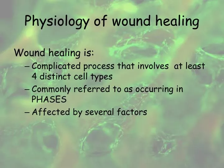
Physiology of wound healing. Wound healing is: Complicated process that involves at least 4 distinct cell types Commonly referred to as occurring in PHASES Affected by several factors. Phases of wound healing process (WHP). Maturation. Proliferation. Inflammation. Haemostasis.

E N D
Physiology of wound healing Wound healing is: • Complicated process that involves at least 4 distinct cell types • Commonly referred to as occurring in PHASES • Affected by several factors
Phases of wound healing process (WHP) Maturation Proliferation Inflammation Haemostasis Where does a chronic wound get stuck?
Platelet Activity WOUND Tissue factor Exposed collagen Extrinsic pathwayVII Intrinsic pathwayXII Messengers for Aggregation & coagulation Intrinsic pathway intermediates IX, VII Platelet Growth factors(PDGF) Coagulation pathway intermediates V, X Other enzymes (proteases) Fibrinogen Prothrombin & thrombin Xlila Cross-linked fibrin clot (structural support for wound healing) FIBRIN
Role of keratinocytes in wound healing Keratinocyte Migration/ Profileration Protease release ECM production Angiogenesis • Dissolves • Nonviable tissue • Fibrin barrier Growth factor/ Cytokine production • Epibloy • Integrins • Chemoattractants • VEGF • KGF (FGF-7) • Matrix formation • Basement membrane formation • VEGF • TGF-α • PDGF • PD-ECGF
Selected growth factors important to wound healing • EGF (epidermal growth factor). Stimulates wound re-epithelialization and stimulates blood vessels and fibroblasts. • FGF (fibroblast growth factor). Stimulates new blood vessel and collagen formation. • PDGF (platelet derived growth factor). Attracts/stimulates smooth muscle cells, fibroblasts, and other cells. Important in ECM formation. • TGF-β(transforming growth factor-beta). Slows buildup of epithelial cells, suppresses immunoglobulin secretion and is helpful in ECM formation. • TNF-a (tumor necrosis factor-alpha). Activates neutrophils, causes fibroblasts to multiply, causes bone/cartilage resorption. • IL-1 (interleukin-1). Attractant for epithelial cells, neutrophils, mono and lymphocytes; also stimulates collagen synthesis.
Chronic wounds – characteristics Increased inflammatory cytokines Altered fibroblast phenotype Abnormalities growth factors Increased proteases Altered keratinocyte function Senescent cells (increased number)
Wound bed preparation • Debridement • Bacterial balance • Dressing therapies (f.i. silver dressings prevent of infections, help reduce healing time)
Local wound care debridement moisture balance Foams Calcium alginates Hydrogels Hydrocolloids Adhesive films Negative pressure therapy Surgical Autolytic Enzymatic Biological
Tissue engineering Biology, medicine and technology are today closely interleaved with each other
Tissue engineering – combining cells and biomaterials into functional tissues Cells are seeded onto a biomaterial scaffold to be integrated into a specific tissue
Tissue engineering Advances • Biological wound dressings • Material scaffolds and cell material interactions • The use of stem cells for tissue engineering • Combination of stem cells and material scaffolds into tissue engineered replacements of tissues and organs
Tissue engineering implants Synthetic polymeric biomaterials • Nonbiodegradable Is required to provide and maintain optimal cellular function -> e.g. alginate, liposome,… • Biodegradable To restore the histological structure and replace the cellular function of recipients -> e.g. poly L-lactic acid, poly glycolic acid, …
Wound healing promoting anti-adhesive matrix The collagen grafting is also applied to produce a healing / promoting antiadhesive membrane • Particularly necessary in peritoneal surgery to prevent postoperative adhesion • To produce skin wound dressing membranes
Methods of tissue bioengineering Skin replacement • Cultured epidermal graft • Cultured human autologous and allogeneic keratinocytes • Semi synthetic materials (composed of human neonatal dermal fibroblasts cultured onto a bioabsorbable mesh)
Methods of tissue bioengineering Active dressings (f.i.: with maggot’s excret, with honey) Photobiomodulation (modulate cellular activity in red to near infrared light) Hyperbaric oxygen therapy: as therapeutic benefit in WT Growth factors (from blood)
Allogeneic cultivated human skin keratinocytes • Make rapid healing of the ulcers particularly those that are difficult to heal • No clinical or laboratory evidence of rejection • No evidence of preexisting cytotoxic antibodies specific fort the HLA class I antigens expressed on HSE cells • A fibrin-based skin substitute produced in the defined keratinocyte medium could be safely used to threat a number of skin defect
Methods of tissue bioengineering • Autologous platelet rich plasma product (platelet gel) • Allogeneic platelet gel The effect is attributed to the growth factors
Impaired healing Delayed unionPseudoarthrosis - nonunion (bone defect) Fracture Impaired healing ? Method of treatment? Infection
Large bone defect Lack of osteogenic progenitor cells • Diabetes, glucocorticoid treatment, chemotherapy, ...
Accepted methods of treatment • Autologous bone transplants • Cancellous bone graft (contains all necessary characteristics of bone substitutes) • Corticocancellous graft (possibly vascularized limited amount) • Homologous (allogeneic) graft Bone banks, treated (no rejection), contains only osteoconductive properties • Ilizarow intercallary bone transport (traction method)
Properties of bone grafts • Osteogenesis (bone marrow, cancellous bone) • Osteoinduction • Demineralized bone matrix • Growth factors (platelet rich plasma, bone morphogenic proteins – BMPs) • Osteoconduction • Ceramics • Collagen
Alternatives Bone substitute (biomaterials for scaffold): • Demineralized bone matrix • Biocompatible ceramics • Synthetic Calcium phosphate • Mineral bone • Collagen • Composite grafts • Osteoinductive collagen
Alternatives Role of GROWTH FACTORS Role of STEM CELLS
Collagen based matrices in tissue engineering Skin equivalent Cartilage repair Bone repair Matrices are also prepared from synthetic polymers
Fracture healing promoting molecules Growth and different factors • The transforming growth factor-β (TGF-β) superfamily • Bone morphogenetic proteins(osteoprogenitors, mesenchymal cells, osteoblasts and chondrocytes within the extracellular matrix produce BMPs.)BMP-2, BMP-4BMP-5, BMP-6, BMP-7GDF-5 (BMP-14), GDF-6 (BMP-13), GDF-7 (BMP-12)BMP-3 (Osteogenin), GDF-10 (BMP-3b) • Platelet-derived growth factor (PDGF) • Fibroblast growth factor (FGFs) • Insulin-like growth factor (IGFs)
Platelet rich plasma (contains high concentrations of growth factors) especially TGF-B and PDGF Autologous Allogeneic PLATELETS MONOCITE NEUTROPHILS PDGF TGF-β FIBROBLAST SMOOTH MUSCLE MACROPHAGE ENDOTHELIUM OSTEOBLASTS PDGF TGF-β
Mesenchymal stem cell: the promise for treating skeletal disorders Adult stem cell are being isolated from various tissues
Adult stem cells Bone marrow contains • Hematopoietic stem cells (HSCs) • All types of blood cells • Bone marrow mesenchymal stem cells (MSCs) • Generating bone, cartilage, fat, fibrous connective tissue
Bone tissue formation Osteogenic progenitor cells Locations: • Periost • Peritrabecular soft tissue • Cancellous bone and bone marrow (in BM aspirate up to 40x less stem cells then in cancellous bone)
Our method of tissue engineering Combined graft Autologous cancellous bone withstem cells Allogeneic platelet gel(source of GFs)
Mixed manually grounded autologous cancellous bone with stem cells corresponding amount of allogeneic platelet concentrate (app. 1,4x10⁹ platelets per 1 ml) AND Added 0,06 ml human thrombin in 40 mMCaCl2 for the activation of platelets in 1 minute the resulting gelled graft can be shaped according to the bone defect and implanted
Our graft Autologous cancellous bone with stem cells and allogeneic platelet gel
Conclusions The use of autologous cancellous bone with stem cells and allogeneic platelet gel is safety and effective method for the treatment of nonunion of long bones
Future Gene arrays for gene discovery
FUTURE Cell and tissue engineering Detection of numerous signal pathways activated during physiological processes Self (re)restoration and differentiation off mammalian embryonic, fetal and stem cells of adult tissues