Download

1 / 38
380 likes | 938 Vues
Brachial Plexus Injuries . Contents. Anatomy of the Brachial Plexus Mechanisms of Brachial Plexus Injury and Pathologies Neurological Evaluation for the Brachial Plexus and Related Special Tests. Paediatric Injuries. History 1764 Obstetrical brachial palsy described by Smellie .

E N D
Contents • Anatomy of the Brachial Plexus • Mechanisms of Brachial Plexus Injury and Pathologies • Neurological Evaluation for the Brachial Plexus and Related Special Tests
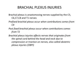
Paediatric Injuries History • 1764 Obstetrical brachial palsy described by Smellie. • 1874 Wilhelm H. Erb described brachial plexus paralysis in adults which involved the upper roots and described certain types of “delivery paralysis”. He credited Duchenne for describing the brachial palsy following delivery in affected newborns. • 1885 Augusta Klumpke first described the clinical picture resulting from injury to lower roots.
Paediatric Injuries • Although injuries can occur at any time, many brachial plexus injuries happen when a baby's shoulders become impacted during delivery and the brachial plexus nerves stretch or tear. • Assoc with: large baby, difficult delivery, gestational diabetes, polyhydramnios, older mum • Incidence = 0.5 to 1.9 per 1000 live births (Bar et al 2001); Brachial plexus palsy occurs in 26% of cases of shoulder dystocia; 90% Erb palsy • Most common on the right side because the most common delivery presentation is left occiput anterior vertex. • Newborns with BP injuries have a higher incidence of low Apgar scores of less than 7 at 1 and 5 mins and of asphyxia than matched controls
Paediatric Injuries • Types of brachial plexus injuries: • avulsion, the most severe type, in which the nerve is torn from the spine • rupture, in which the nerve is torn but not at the spinal attachment • neuroma, in which the nerve has torn and healed but scar tissue puts pressure on the injured nerve and prevents it from conducting signals to the muscles • neuropraxia or stretch, in which the nerve has been damaged but not torn; most common type of brachial plexus injury • Treatment: • Conservative: Many children who are injured during birth improve or recover by 3 to 4 months of age. Treatment for brachial plexus injuries includes physical therapy and, in some cases, surgery. • Prognosis: The site and type of brachial plexus injury determines the prognosis. For avulsion and rupture injuries, there is no potential for recovery unless surgical reconnection is made in a timely manner. The potential for recovery varies for neuroma and neuropraxia injuries. Most individuals with neuropraxia injuries recover spontaneously with a 90-100% return of function.
Paediatric Injuries What is your management of the obstetric brachial plexus injury? • History: Large baby; difficult delivery; shoulder distocia; maternal DM • Examination: try to determine level • At bith: look at upper limb posture • At 3/12: look for elbow flexion (sign of recovery) • When older ask them to take off shirt and watch • Investigations: MRI, myelogram • Management: • Physio: passive stretch; maitain FPROM; prevent contractures • Surgery at 3/12 (20%): explore neck via L-shaped incision in posterior triangle; nerve graft vsneurolysis (give preference to lower roots, so they develop hand and elbow function) • Surgery at 8 yrs: tendon transfers
Observe Posture and Movements • Baby will just lie there! Ask mum if both arms move / twitch. Dangle some keys to see if it will reach out? Baby will just lie there! Ask mum if both arms move / twitch. Dangle some keys to see if it will reach out? This child can not reach up!
Observe Posture and Movements • Get child to undress and see how he gets on • Comment on what u see!
Brachial Plexus Injury: Adults • High-energy trauma to the upper extremity and neck causes a variety of lesions to the brachial plexus. • The common mechanism is violent distraction of the entire forequarter from the rest of the body ie motorcycle accident or a high-speed motor vehicle accident. A fall from a significant height may also result in brachial plexus injury. • Sports most commonly associated with brachial plexus injuries include: Am football, baseball, basketball, volleyball, fencing, wrestling, and gymnastics • Nerve injuries can result from blunt force trauma, poor posture, or chronic repetitive stress • Patients generally present with pain and/or muscle weakness • Over time, some patients may experience muscle atrophy • Loss of useful function of the upper extremity is common
Mechanisms of Injury to the Brachial Plexus • Traction: direct blow to the shoulder with the neck laterally flexed toward the unaffected shoulder (gymnast falls on beam) • Direct trauma: direct blow to the supraclavicularfossa over Erb’s point • C. Cervical Nerve Compression: Occurs when the neck is flexed laterally toward the patient’s affected shoulder, compressing / irritating the nerves, resulting in point tenderness over involved vertebrae of affected nerve(s) • (Troub, 2001)
Injury Classification Millesi classification* • Supraganglionic • Infraganglionic • Trunk • Cord Anatomical Classification • C5-6 waiters tip (Erbs palsy) • C5-7 as above, elbow slightly flexed • C5-T1 flail limb, claw hand, vasomotor changes, +/- Horners syndrome
Grades of Injury • Grade 1 – Neuropraxia • Disruption in nerve function that produces numbness and tingling • Most common grade within athletics • Symptoms usually resolve within several minutes • Grade 2 – Axonotmesis • Damage to the nerve’s axon • Symptoms = numbness, tingling, and affected function (may last several days) • Long nerves have a greater healing time than short nerves • Rare within athletics • Grade 3 – Neurotmesis • Permanent nerve damage occurs • Very rare within athletics • “Occurs with high-energy trauma, fractures, and penetrating injuries”
Adult Brachial Plexus Injury How do you Rx the patient knocked off his motorcycle with clavicle # and flail arm? • Manage acute injury according to ATLS principles; look for concomitant injury iec-spine. • History • Age, handedness, occupation, special skills • Cause of injury: arm hyperabductedvs neck laterally flexed • Immediate or delayed arm weakness • Concomitant injury • General health: PMH, DH, Smoker
Adult Brachial Plexus Injury Examination (use pre-printed brachial plexus diagrams): determine level • Look at face: does he have Horner’s? (=lower root lesion C8 T1) • Undress upper torso • Look from front at posture of arm, scars, muscle wasting, asymmetry/swelling • Look at back again for scars, muscle wasting, asymmetry • Test sp. Accessory n (shrug shoulders) • Supraspinatus responsible for 1st 20 of shoulder abduction (resisted arm abduction) • Rhomboids (touch back of head) • Lat dorsi (press both hands into hips and cough) • Look at vascularity of arm • Check sensation both upper limbs (root levels) • Check movement both upper limbs from shoulder to fingers (AROM + PROM) • Reflexes • Function of phrenic nerve
Examination • LOOK, FEEL, MOVE (Talk as you are doing) • Manage according to EMST/ATLS in acute setting. Abrasions to the head, helmet, or tip of the shoulder suggest supraclavicular injury. • Look at the face: Ptosis, myosis and anhydrosis (Horner syndrome) suggest a complete lower plexus lesion
Examine the Back Wall test for serratus ant (winging scapula) Note weak trapezius (asymmetric shrug)
Examine the Back • Swelling about the shoulder can be dramatic. Diminished or absent pulses suggest vascular injury, and special consideration should be given to rupture of the subclavian vessels. Clavicle fractures are often palpable. Careful inspection and palpation of the axial skeleton may reveal concomitant injuries. Examine each cervical root individually for motor and sensory function as soon as circumstances allow.
Examine the Back Photograph showing patient with left shoulder subluxation resulting from a flail arm caused by C5–T1 lesions. Note the left deltoid, supra-, and infraspinatus muscle atrophy
Examine the Back Is lat dorsi present?
Examine Front Notice clavicular scar, posture, wasting of Deltoid and biceps
Examine Front • If you see a flap mention it and look for the donor site!
Related Special Tests Brachial Plexus • Cervical Compression Test • Cervical Distraction Test • Spurling’s Test • Brachial Plexus Traction Test Thoracic Outlet Syndrome • Adson’s Test • Allen’s Test • Military Brace Position
Peripheral Nerve Tests Axillary N. • Sensory – Lateral arm • Motor – Shoulder abduction Musculocutaneous N. • Sensory – Anterior arm • Motor – Elbow flexion Radial N. • Sensory – 1st Dorsal web space • Motor – Wrist extension and thumb extension Median N. • Sensory – Pad of Index finger • Motor – Thumb pinch and abduction Ulnar N. • Sensory – Pad of little finger • Motor – Finger abduction
Reflex Tests • C5 – Biceps brachii reflex (anterior arm near antecubitalfossa) • C6 – Brachioradialis reflex (lateral aspect of forearm) • C7 – Triceps brachii reflex (at insertion of tricepbrachii) • C8 and T1 do not have reflex tests
Investigations Imaging: Xray: AP chest (look for teeth and fractures ), AP + lat views shoulder, C-Spine (AP, lat, odontoid peg), Fine-cut CT, MRI
Investigations • Sensory nerve action potentials (SNAPs): differentiate preganglionic from postganglionic injuries. • Electromyography (EMG): In the first week after injury, EMG cannot be used to exclude a complete disruption unless voluntary motor unit action potentials are observed. If no signs of denervation are present in a paralyzed muscle by 3 weeks after injury, EMG can be used to confirm a neuropraxia. • Somatosensory evoked potentials (SSEPs): In general, SNAPs are more reliable than SSEPs. Many difficulties exist with SSEPs, and they are not widely used.
Management • Medical: MDT • physio: maintain supple joints with FROM • Orthoptists / splinting • Pain control • Surgical options: • nerve transfers • nerve grafting • muscle transfers • free muscle transfers • neurolysis of scar in incomplete lesions • Arthrodesis to stabilise joints
Management Surgical options: Immediate vs delayed (timing contraversial) • Indications for Surgery at time of injury • Open injury • High energy injury • Supraclavicular injury • Associated depressed clavicle fracture :explore and immediate repair / nerve grafts • Surgery 3/12 post injury IF CLOSED (and no sign recovery): nerve grafts (if not done B4); nerve transfer if supraganglionic • Surgery >1 year post injury: local or free muscle transfer starting at proximal joint (eg 2-stage reconstruction with sural nerve cross-thorax graft, attached to nerve to pec minor or long thoracic, then free contralat LD)
Planning for Reconstruction • What is the loss? • What is the need? • What is possible? • What is available? • What are the other injuries? • Is later surgery needed and what can be done now?
What is the loss? • Shoulder motion • Shoulder stability • Elbow flexion • Wrist and hand function • Sensation • Pain • Trophic changes • Body image
What is available? • Primary repair: Very rare • Neurolysis only with late surgery • Plexus anatomical cable grafting • Nerve transfers • Accessory nerve • Cervical plexus • Phrenic nerve • Intercostal nerves • Ulnar ECU nerve • Crossed C7 • Hypoglossal nerve • Nerve grafts • Sural • medial cutaneous forearm • ulnar (vascularised)