Download
1 / 51
560 likes | 820 Vues
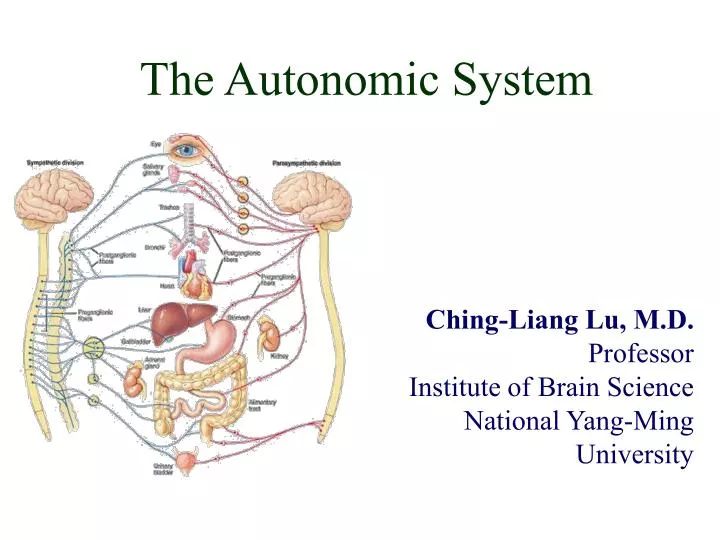
The Autonomic System. Ching-Liang Lu, M.D. Professor Institute of Brain Science National Yang-Ming University. Autonomic Nervous System (ANS). Innervate smooth and cardiac muscle and glands Make adjustments to ensure optimal support for body activities Operate via subconscious control

E N D
The Autonomic System Ching-Liang Lu, M.D. Professor Institute of Brain Science National Yang-Ming University
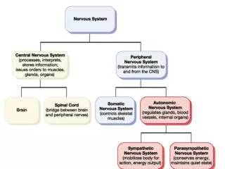
Autonomic Nervous System (ANS) • Innervate smooth and cardiac muscle and glands • Make adjustments to ensure optimal support for body activities • Operate via subconscious control • Have viscera as most of their effectors
Sensory/Motor + Somatic/Visceral Somatic Nervous System Autonomic Nervous System
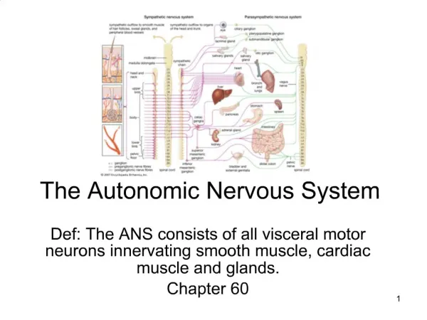
Divisions of the ANS • Sympathetic division (thoracolumbar,“fight or flight”) • Thoracic and lumbar segments • Parasympathetic division (craniosacral, “rest and repose”) • Preganglionic fibers leaving the brain and sacral segments • Enteric nervous system (ENS) • May work independently
Sympathetic and Parasympathetic • Often they have opposing effects • May work independently • May work together each one controlling one stage of the process
Overview of ANS Functional Differences Sympathetic • “Fight or flight” • Catabolic (expend energy) Parasympathetic • “Feed & breed”, “rest & digest” • Homeostasis » Dual innervation of many organs — having a brake and an accelerator provides more control
Somatic vs. AutonomicNervous Systems • The ANS differs from the SNS in the following three areas • Effectors • Efferent pathways • Target organ responses
Somatic vs. Autonomic Systems:Effector • The effectors of the SNS are skeletal muscles • The effectors of the ANS are cardiac muscle, smooth muscle, and glands
Somatic vs. Autonomic Systems: Efferent Pathways • Heavily myelinated axons of the somatic motor neurons extend from the CNS to the effector • Axons of the ANS are a two-neuron chain • The preganglionic (first) neuron has a lightly myelinated axon • The ganglionic (second) neuron extends to an effector organ
Overview of the Autonomic Nervous System Similarities between Sympathetic & Parasympathetic • Both are efferent (motor) systems: “visceromotor” • Both involve regulation of the“internal”environment generally outside of our conscious control: “autonomous” • Both involve 2 neurons that synapse in a peripheral ganglion • Innervate glands, smooth muscle, cardiac muscle glands ganglion CNS smooth muscle cardiac muscle preganglionic neuron postganglionic neuron
Overview of the Autonomic Nervous System Differences between Sympathetic & Parasympathetic Location of Preganglionic Cell Bodies Sympathetic Parasympathetic Thoracolumbar T1 – L2/L3 levels of the spinal cord Craniosacral Brain: CN III, VII, IX, X Spinal cord: S2 – S4
Overview of the Autonomic Nervous System Differences between Sympathetic & Parasympathetic Relative Lengths of Neurons Sympathetic target ganglion CNS short preganglionic neuron long postganglionic neuron Parasympathetic target ganglion CNS long preganglionic neuron short postganglionic neuron
Overview of the Autonomic Nervous System Differences between Sympathetic & Parasympathetic Neurotransmitters Sympathetic NE (ACh at sweat glands), + / -, α & ß receptors ACh, + • All preganglionics release acetylcholine (ACh) & are excitatory (+) • Symp. postgangl. — norepinephrine (NE) & are excitatory (+) or inhibitory (-) • Parasymp. postgangl. — ACh & are excitatory (+) or inhibitory (-) • Excitation or inhibition is a receptor-dependent & receptor-mediated response ACh, + Parasympathetic Potential for pharmacologic modulation of autonomic responses ACh, + / - muscarinic receptors
Overview of the Autonomic Nervous System Differences between Sympathetic & Parasympathetic Target Tissues Sympathetic Parasympathetic • Organs of head, neck, trunk, & external genitalia • Organs of head, neck, trunk, & external genitalia • Adrenal medulla • Sweat glands in skin • Arrector muscles of hair • ALL vascular smooth muscle » Sympathetic system is distributed to essentially all tissues (because of vascular smooth muscle) » Parasympathetic system never reaches limbs or body wall (except for external genitalia)
Sympathetic division anatomy • Preganglionic neurons between segments T1 and L2 – lateral gray horn of spinal cord • Preganglionic fibers • Short • Travel in the ventral root and spinal nerve • Ganglionic neurons in ganglia near vertebral column • Specialized neurons in adrenal glands • Postganglionic fibers • Long fibers
Sympathetic ganglia • Sympathetic chain ganglia(paravertebral ganglia) • Typically there are 23 ganglia – 3 cervical, 11 thoracic, 4 lumbar, 4 sacral,and 1 coccygeal • Collateral ganglia (prevertebral ganglia) • Adrenal medulla
Structure of spinal nerves: Somatic pathways dorsal ramus dorsal root ganglion dorsal root spinal nerve somatic sensory nerve (GSA) dorsal horn CNS inter- neuron somatic motor nerve (GSE) ventral horn ventral ramus gray ramus communicans ventral root white ramus communicans Mixed Spinal Nerve sympathetic ganglion
Structure of spinal nerves: Sympathetic pathways dorsal ramus intermediolateral gray column spinal nerve ventral ramus gray ramus communicans white ramus communicans sympathetic ganglion
Organization and anatomy of the sympathetic division • Segments T1-L2, ventral roots give rise to myelinated white ramus • Leads to sympathetic chain ganglia
Postganglionic fibers of thesympathetic ganglia • Some fibers will return to the spinal nerve through a gray ramus and will innervate skin, blood vessels, sweat glands, adipose tissue, arrector pili muscle • Some fibers will form sympathetic nerves that will innervate thoracic organs • Go directly to innervate the thoracic organs
Sympathetic System: Postganglionic Cell Bodies 1. Paravertebral ganglia • Located along sides of vertebrae • United by preganglionics into Sympathetic Trunk • Preganglionic neurons are thoracolumbar (T1–L2/L3) but postganglionic neurons are cervical to coccyx • Some preganglionics ascend or descend in trunk Paravertebral ganglia sympathetic trunk (chain) synapse at same level Prevertebral ganglia • celiac ganglion • sup. mesent. g. • inf. mesent. g. ascend to synapse at higher level descend to synapse at lower level aorta Moore’s COA5 2006
Collateral (prevertebra) ganglia • Preganglionic fibers will pass through the sympathetic chain without synapsing • Preganglionic fibers will synapse within collateral ganglia (prevertebra ganglia) • Splanchnic nerves will synapse on one of the 4 collateral ganglions
Collateral (prevertebra) ganglia • Celiac ganglion • Postganglionic fibers innervates stomach, liver, gall bladder, pancreas, spleen • Superior mesenteric ganglion • Posganglinic fibers innervates small intestine and initial portion of large intestine • Inferior mesenteric ganglion • Postganglionic fibers innervate the final portion of large intestine • Inferior hypogastric • Posganglionic fibers innervates urinary bladder , sex organs
Sympathetic System: Postganglionic Cell Bodies 2. Prevertebral (preaortic) ganglia • Located anterior to abdominal aorta, in plexuses surrounding its major branches • Preganglionics reach prevertebral ganglia via abdominopelvic splanchnic nerves Paravertebral ganglia sympathetic trunk (chain) • Prevertebral ganglia • • celiac ganglion • • sup. mesent. g. • • inf. mesent. g. • inf. hypogastric abdominopelvic splanchnic nerve aorta Moore’s COA5 2006
Adrenal medulla • Preganglionic fibers will pass through sympathetic ganglia without synapsing • Preganglionic fibers will synapse on adrenal medulla • Adrenal medulla will secrete • Epinephrine • Norepinephrine
Adrenal medulla • Neurotransmitter will go into general circulation • Their effects last longer than those produced by direct sympathetic innervation
Adrenal gland is exception • Synapse in gland • Can cause body-wide release of epinephrine (adernalin) and norepinephrine in an extreme emergency (adrenaline “rush” or surge)
Sympathetic System: Summary visceral tissues (organs) Cardiopulmonary Splanchnics: postganglionic fibers to thoracic viscera somatic tissues (body wall, limbs) T1 postganglionics via 31 spinal nerves to somatic tissues of neck, body wall, and limbs Abdominopelvic Splanchnics: preganglionic fibers to prevertebral ganglia, postganglionic fibers to abdominopelvic viscera sympathetic trunk L2 prevertebral ganglia Moore’s COA5 2006
Role of the Sympathetic Division • The sympathetic division is the “fightor-flight” system • Involves E activities – exercise, excitement, emergency, and embarrassment • Promotes adjustments during exercise– blood flow to organs is reduced, flow to muscles is increased
Role of the Sympathetic Division • Its activity is illustrated by a person who is threatened • Heart rate increases, and breathing is rapid and deep • The skin is cold and sweaty, and the pupils dilate
Parasympathetic division(craniosacral division) • Preganglionic neurons in the brainstem(nuclei of cranial nerves III, VII, IX, X) and sacral segments of spinal cord (S2-S4) • Ganglionic neurons in peripheral ganglia located within or near target Organs • Terminal ganglion • Intramural ganglion
Parasympathetic Pathways Cranial outflow • CN III, VII, IX, X • Four ganglia in head • Vagus nerve (CN X) is major preganglionic parasymp. supply to thorax & abdomen • Synapse in ganglia within wall of the target organs (e.g., enteric plexus of GI tract) Sacral outflow • S2–S4 via pelvic splanchnics • Hindgut, pelvic viscera, and external genitalia Clinical Relevance » Surgery for colorectal cancer puts pelvic splanchnics at risk » Damage causes bladder & sexual dysfunction Moore’s COA5 2006
Parasympathetic activation • Effects produced by the parasympathetic division • Relaxation • food processing • energy absorption • Pupil constriction • Constriction of respiratory passageway • Decrease heart rate and blood pressure • Stimulates defecation and urination
Referred Pain • Pain stimuli arising from the viscera are perceived as somatic in origin • This may be due to the fact that visceral pain afferents travel along the same pathways as somatic pain fibers
Visceral Afferents and Referred Pain dorsal root ganglion Visceral sensory nerves [GVA] • run with sympathetic nerves • cell bodies in dorsal root ganglion • nerve ending in viscera Somatic sensation: • conscious, sharp, well-localized • touch, pain, temperature, pressure, proprioception Visceral sensation: • often unconscious; if conscious: dull, poorly-localized • distension, blood gas, blood pressure, cramping, irritants
Visceral Afferents and Referred Pain Referred Pain: • Pain originating in a visceral structure perceived as being from an area of skin innervated by the same segmental level as the visceral afferent • Results from convergence of somatic & visceral afferents on the same segmental level of the spinal cord • “Cross-talk” in the dorsal horn convergence & “cross-talk” somatic afferent www.merck.com visceral afferent Kandel et al. 2000
Visceral Afferents and Referred Pain Maps of Referred Pain Grant’s Atlas 11 2005
Interactions of the AutonomicDivisions • Most visceral organs are dual-innervated • both sympathetic and parasympathetic fibers • dynamic antagonisms that precisely control visceral activity • Sympathetic fibers increase heart and respiratory rates, and inhibit digestion and elimination. • Parasympathetic fibers decrease heart and respiratory rates, and allow for digestion and the discarding of wastes
Cooperative Effects • Example: control of external genitalia • Parasympathetic fibers: • vasodilation erection of the penis and clitoris • Sympathetic fibers • cause ejaculation of semen in males and reflex contraction of a female vagina
Unique Roles of the Sympathetic Division • Regulates many functions not subject to parasympathetic influence • These include the activity of the adrenal medulla, sweat glands, arrector pili muscles, kidneys, and most blood vessels • The sympathetic division controls: • Thermoregulatory responses to heat • Release of renin from the kidneys • Metabolic effects • Raises blood glucose levels • Mobilizes fat as a food source • Stimulates the reticular activating system (RAS) of the brain, increasing mental alertness
Localized Versus Diffuse Effects • The parasympathetic division exerts short-lived, highly localized control • The sympathetic division exerts long-lasting, diffuse effects
Central control of the Autonomic NS Amygdala:main limbic region for emotions -Stimulates sympathetic activity, especially previously learned fear-related behavior -Can be voluntary when decide to recall frightful experience - cerebral cortex acts through amygdala -Some people can regulate some autonomic activities by gaining extraordinary control over their emotions Hypothalamus: main integration center Reticular formation: most direct influence over autonomic function
Hypothalamic Control • Centers of the hypothalamus control: • Heart activity and blood pressure • Body temperature, water balance,and endocrine activity • Emotional stages (rage, pleasure) and biological drives (hunger, thirst, sex) • Reactions to “fear” and the “fight or-flight” system
Neural innervation of bowel • Autonomic nervous system • Extrinsic set of nerves • Parasympathetic • Sympathetic • Enteric nervous system (ENS) • Intrinsic set of nerves • ~108 neurons - similar to spinal cord “brain of gut” • Neurons extending from esophagus to anus • 2 plexuses • Myenteric plexus • Submucosal plexus
Intrinsic Nervous System • Myenteric plexus (Auerbach) • Located between the longitudinal and circular layers of muscle in the tunica muscularis • Controls tonic and rhythmic contractions • Exerts control primarily over digestive tract motility • Submucosal plexus (Meissner) • Buried in the submucosa • Senses the environment within the lumen • Regulates GI blood flow • Controls epithelial cell function (local intestinal secretion and absorption) • May be sparse or missing in some parts of GI tract
Intrinsic Nervous System • 3 types of neurons in enteric system • Sensory neurons (5 types) • Chemoreceptors sensitive to acid, glucose and amino acids have been demonstrated which, in essence, allows "tasting" of lumenal contents. Sensory receptors in muscle respond to stretch and tension • Motor neurons • Control GI motility and secretion, and possibly absorption • Interneurons • Largely responsible for integrating information from sensory neurons and providing it to motor neurons