Download
1 / 23
230 likes | 368 Vues
International Symposium AT University of Mumbai 9 th March 2012 “Histopathological changes in tissues of Dosinia fibula exposed to WSFs of crude oilâ€. Prin. Dr. (Mrs.) Snehal S. Donde. MSc, PhD, PGDEM, MBA HIND SEVA PARISAD’S PUBLIC NIGHT DEGREE COLLEGE

E N D
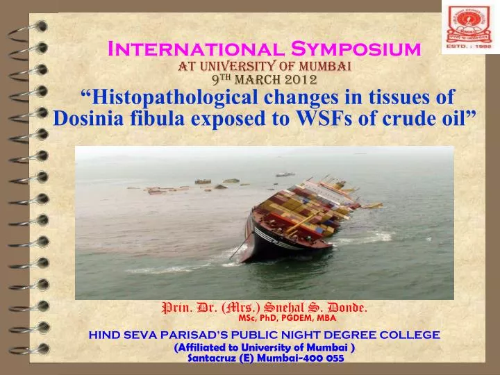
International SymposiumAT University of Mumbai 9th March 2012“Histopathological changes in tissues of Dosinia fibula exposed to WSFs of crude oil” Prin. Dr. (Mrs.) Snehal S. Donde. MSc, PhD, PGDEM, MBA HIND SEVA PARISAD’S PUBLIC NIGHT DEGREE COLLEGE (Affiliated to University of Mumbai ) Santacruz (E) Mumbai-400 055
Inevitable oil spills • Two Panamanian cargo ships - MSC Chitra and the MV Khalijia-III collided off the Mumbai coast, triggering an oil spill, on August 7, 2010-800 tonnes • Mumbai-Uran pipeline spill- January 21, 2011-55 tones • The oil slick as seen from space by NASA’s Terra satellite on May 24, 2010 Location Gulf of Mexico near • Disposal of Ballast water
Objectives of study Discuss histopathological alterations in different tissues of soft shelled clam Dosiniafibula ( Reeve) due to toxic effect of WSF of BH crude oil Crude oil pollution needs greater attention as it is complex mixture of aliphatic, acyclic and aromatic hydrocarbons. Aromatic hydrocarbons are tainting components and exhibit carcinogenic effect (Rice et.al.1977) and are potentially toxic because they are relatively soluble in water (Siron et.al.1987). Higher concentration of the pollutants cause histological changes and also collapses the biochemical composition.
Materials and methods • Clams (length 25-30mm) acclimatized for 7days and were kept in filtered sea water during experimental period of 120 hrs. • A series of stock WSFs were prepared (10%, 20%, and 30 % concentrations by v/v oil-water mixing ) by proportional oil and sea water mixing at constant temperature (Neff and Anderson,1977). • Clams exposed to sublethal concentrations of 5, 10, 25, 50, 75 and 100 percent WSF dilution concentrations were sacrificed after intervals of every 7 days for 28 days and morphological alterations in the gills, hepatopancreas (digestive diverticula), mantle and adductor muscles were studied. • After each exposure period the tissues were fixed in Bouin's fixative and the tissues were processed as per the routine microtechniques methods and the sections (7 to 8 μm thick) were cut and stained with eosin and Ehrlich's hematoxylin for further studies.
Shell thickness • Gafrarium divaricatum- upper (0.250 + 0.11 cm), middle (0.272 + 0.09 cm) and lower (0.305 + 0.01 cm) • Dosinia fibula upper (0.228 + 0.03), middle (0.161 +0.01cm) and lower (0.152 + 0.10 cm).
LC50 • LC50 values of WSF (ppb) of PG crude oil • 48, 72, 96 and 120hrs readings recorded
Relative potency and parallelism between Gafrarium divaricatum and Dodiniafibula toWSFs of PG crude oil. (Values are mean + SD of five determinations) • SR < fSR hence curves are parallel within experimental error. • PR > fPR the two compared differ significantly in their potency
Accumulation and depuration of PAH in two species of marine bivalve in 10% WSF stock ( g / g ) (Values are mean + SD of five determinations ) PH- 7.5+1.5 ; Dissolved Oxygen - 5.8+ 0.9 mg / l ; Temperature - 27 + 2 o C ; Salinity - 30.2+ 1.05 %o *P < 0.01 **P <0.05.
The relation of oil accumulation to the wet wt and lipid content of Gafrarium divaricatum and Dosinia fibula during 96 hr of exposure
Plate no: 1.1- Normal gill of control (7th day): showing cavity between the lamellae divided by several partitions, the interlamellar junctions (ILJ), and a number of vertical compartments called water tubes (WT). Filled blood sinuses (BS) are very promenient. Plate no: 1.2 Exposed gill of 5% WSF dilution (7th day): showing incomplete interlamellar junctions (ILJ), Widening of water tubes (WWT), Necrotic junction (NJ) and loss of epithelium (LE). Vacuolization and desquamation in lamellae is seen.
Plate: 1.25 general loss of structural integrity . Activities of eulaterofrontal cirri, necrosis of gill epithelium cells, Loss of cilia and epithelial cells from filaments reduce filtering efficiency. Rupture the hemolymph sinuses it will lead to the loss of hemolymph • Plate: 1.26 damaged ciliary tufts, Tightly meshed gill structure to prevent finest sediment from entering the suprabrabchial cavity can be seen. showing damaged lamellar junctions widening of water tube and ostium.
Plate no: 2.1 - Magnification 100X) Normal digestive diverticula of control (7th day): Showing folding in epithelium (FE), inner lumenar space (ILS) and basement membrane (BM). Plate no: 2.5 - (Magnification 100X)Exposed digestive diverticula of 50% WSF dilution (7th day): showing loss of cilia (LC) in border of typhlosole, disruption of inner lining of tubule (DLT), and infiltration of haemocytes (IH). Atrophy of diverticula, disintegration of epithelial cells, necrotic epithelium with sloughing and vacuolization (VE). Reduced lumenar space Loss of ciliated border of typhlosole Infiltration of haemocytes
Plate: 2.13 -Digestive diverticula of 100% WSF dilution (7th day): secondary tubule showing mixing of cellular content of different tubules, Fusion of nuclei, infiltration of haemocytes, detachment of epithelial cells from basement membrane, loss of typhlosolePlate: 2.14- Digestive diverticula of 50% WSF dilution (7th day):showing loss of brush border (LC) and disintegration of inner layer of epithelial cells. showing vacuolization (hyperplasia and cellular proliferation) of epithelial cells (VE) and absence of bubbling cells and partial disintegration. Sloughing of epithelium affected the function of removal of metabolic wastes
Plate no: 3.1 - (Magnification 100X) Normal mantle of control (7th day): section showing outer columnar epithelium (OE), middle fibrous connective tissue (FCT) and inner highly folded ciliated epithelium (ICE). outer columnar epithelium • Plate no: 3.2 - (Magnification 100X)Exposed mantle of 5% WSF dilution (28th day): showing loss of basal membrane (LBA) and damage to fibrous connective tissue, disintegration of mucus sereting cells, total loss of structural integrity, disruption of hemocoelic spaces, and intermingled cells. • Inner highly folded epithelial cells
Plate: 3.3- Exposed mantle of 25% WSF dilution (7th day):shows reduction in haemocoelic spaces (SIA) and shrinking of epithelia (SE). It secrete shell and is an important supplementary respiratory organ • Plate: 3.4- Exposed mantle of 50% WSF dilution (7th day): showing vacuolization of epithelial cell (VEC), damage caused to epithelial cells (DEC), desquamation of connective tissue (DCT) and disintegration of mucus secreting cells (MSC). Disintegration of mucus secreting cells
Plate no: 4.1 - (Magnification 100X) Normal adductor muscle of control(7th day): showing striated and smooth muscle fibres with extended inter muscular spaces (EIMS) structural integrity of the tissue is seen Plate no: 4.2 - (Magnification 100X) Exposed adductor muscle of 5% WSF dilution (7th day): section showing reduced intermuscular space (RIMS) and few necrotic fibres (LST). wide gap between the inter fibrous spaces, infiltration of hemocytes near thick band of tissues
Plate: 4.11- Adductor muscle of 5% WSF dilution (28th day): showing disintegration of striated (SF) and smooth muscle fibes (SM). Adductor muscles play an important role in all kinds of muscular action upon the contents of the mantle cavity. • Plate: 4.12- Aadductor muscle of 10% WSF dilution (28th day): showing disrupted fibres (SD) and total loss of structural integration.
Summary & Conclusion • Bivalve molluscs in their natural environment are frequently exposed to single spill or chronic discharges of petroleum oil. • Marine bivalves accumulate petroleum hydrocarbon in their tissues. • In clams, the gills, digestive diverticula have been identified as excellent biological indicators of the effects of toxic materials in the ambient environment. • Physiological functions are largely dependent on structure and morphological changes. • The structural damage in gills thus leads to reduced respiration rate, clearance rate (sluggish activities of cirri) and general loss of regulatory mechanism. • In the digestive gland a loss of synchronization of intracellular digestion between individual tubules affect both absorption and secretion, disturb the detoxication mechanism. • Mantle secrete shell and is an important supplementary respiratory organ, hence any damage cause retarded shell growth and affect respiration. • The adductors facilitate sustained and rapid closing of the valves and any damage affect flushing out.
Cont---- • Siphons are important renewable food, disintegrated muscles of the mantle affected the contraction of siphons. Also drawing of water towards gill gets affected and may lead to starvation and low oxygen consumption. • The damage caused to mucus glands situated in the mantle, rendered the body of the clam to be in direct contact with the pollutant. • Water is expelled from the mantle cavity by sudden contractions of adductor muscles in order to flush out accumulations of pseudofaeces
Future strategy • The research study was conducted in cooperation of NIO & CIFE. • Research project with awareness programme are further planned as follows: • Conducting regular surveys. • Public awareness programs. • Collaborative work in direction of bioremediation. (Microbially mediated removal of contaminants) • Research in efficacy and effects of Oil spill dispersants • Publication of data
References: • Anderson, J.W., J.M. Neff, B.A. Cox, H.E. Tatem, G.M.Hightower 1974. Mar.Biol. 27,75-88. • Cohen. 1997. Environ. Pollut. 12: 173-189. • Moles,A. 1998. Environ. Contam. Toxicol. 61: 102-107. • Metcalf, J.L., M.N. Carlton 1990.Sci. Tot. Environ. 97/98: 595-615. • Neff, J.M., B.A. Cox, D. Dixit and J.W. Anderson 1976. Mar. Biol. 38(3): 279-289. • Neff,J.M. and J.W. Anderson 1975.In 1975 oil spill conference proceedings. American Petroleum Institute. Washington D.C. pp. 469-472. • Rice, S.D., D.A. Moles, T.L. Taylor, J.I. Karinen 1979. In Proceeding of the 1979 oil spill conference, American Petroleum Institute. • Shaw,G.David, Thomas.E.Hogan, Douglas, J. McIntosh 1986. Estuarine. Coastaland Shelf Science. 23: 863-872. • Stainken, D.M. 1976. Environ. Cont. Toxicol. 16: 730-738. • Stegeman, J.J. and J.M. Teal 1973. Mar. Biol. 22: 37-44. • marinebiotech.org • http://edac_ecowatch.northerngulfinstitute.org/activity/survey.html (my paper) • http://www.nap.edu/catalog.php?record_id=11283 • http://wiki.ask.com/Deepwater_Horizon_oil_spill